Show PDB file:   
         Plain Text   HTML   (compressed file size)
QuickSearch:   
by PDB,NDB,UniProt,PROSITE Code or Search Term(s)  
(-)Asym.Unit - manually
(-)Asymmetric Unit
(-)Asym. Unit - sites
(-)Biological Unit 1
(-)Biol. Unit 1 - sites
(-)Biological Unit 2
collapse expand < >
Image Asym.Unit - manually
Asym.Unit - manually  (Jmol Viewer)
Image Asymmetric Unit
Asymmetric Unit  (Jmol Viewer)
Image Asym. Unit - sites
Asym. Unit - sites  (Jmol Viewer)
Image Biological Unit 1
Biological Unit 1  (Jmol Viewer)
Image Biol. Unit 1 - sites
Biol. Unit 1 - sites  (Jmol Viewer)
Image Biological Unit 2
Biological Unit 2  (Jmol Viewer)

(-) Description

Title :  STRUCTURE OF PROTEIN OF UNKNOWN FUNCTION PA5198 FROM PSEUDOMONAS AERUGINOSA
 
Authors :  J. Osipiuk, E. Evdokimova, A. Savchenko, A. Edwards, A. Joachimiak, Mi Center For Structural Genomics (Mcsg)
Date :  04 May 05  (Deposition) - 21 Jun 05  (Release) - 13 Jul 11  (Revision)
Method :  X-RAY DIFFRACTION
Resolution :  1.10
Chains :  Asym. Unit :  A,B
Biol. Unit 1:  A  (1x)
Biol. Unit 2:  B  (1x)
Keywords :  Structural Genomics, Hypothetical Protein, Pa5198, Psi, Protein Structure Initiative, Midwest Center For Structural Genomics, Mcsg, Unknown Function (Keyword Search: [Gene Ontology, PubMed, Web (Google))
 
Reference :  J. Osipiuk, E. Evdokimova, A. Savchenko, A. Edwards, A. Joachimiak
X-Ray Crystal Structure Of Hypothetical Protein Pa5198 At 1. 1 A Resolution.
To Be Published
PubMed: search

(-) Compounds

Molecule 1 - HYPOTHETICAL PROTEIN PA5198
    ChainsA, B
    EngineeredYES
    Expression SystemESCHERICHIA COLI BL21(DE3)
    Expression System PlasmidPET15B
    Expression System StrainBL21(DE3)
    Expression System Taxid469008
    Expression System Vector TypePLASMID
    GenePA5198
    Organism ScientificPSEUDOMONAS AERUGINOSA
    Organism Taxid287

 Structural Features

(-) Chains, Units

  12
Asymmetric Unit AB
Biological Unit 1 (1x)A 
Biological Unit 2 (1x) B

Summary Information (see also Sequences/Alignments below)

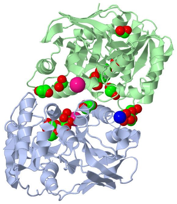
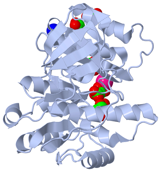
(-) Ligands, Modified Residues, Ions  (6, 18)

Asymmetric Unit (6, 18)
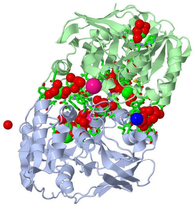
No.NameCountTypeFull Name
1EDO12Ligand/Ion1,2-ETHANEDIOL
2GOL1Ligand/IonGLYCEROL
3K2Ligand/IonPOTASSIUM ION
4NA1Ligand/IonSODIUM ION
5PEG1Ligand/IonDI(HYDROXYETHYL)ETHER
6TLA1Ligand/IonL(+)-TARTARIC ACID
Biological Unit 1 (3, 6)
No.NameCountTypeFull Name
1EDO4Ligand/Ion1,2-ETHANEDIOL
2GOL1Ligand/IonGLYCEROL
3K-1Ligand/IonPOTASSIUM ION
4NA-1Ligand/IonSODIUM ION
5PEG-1Ligand/IonDI(HYDROXYETHYL)ETHER
6TLA1Ligand/IonL(+)-TARTARIC ACID
Biological Unit 2 (2, 9)
No.NameCountTypeFull Name
1EDO8Ligand/Ion1,2-ETHANEDIOL
2GOL-1Ligand/IonGLYCEROL
3K-1Ligand/IonPOTASSIUM ION
4NA-1Ligand/IonSODIUM ION
5PEG1Ligand/IonDI(HYDROXYETHYL)ETHER
6TLA-1Ligand/IonL(+)-TARTARIC ACID

(-) Sites  (18, 18)

Asymmetric Unit (18, 18)
No.NameEvidenceResiduesDescription
01AC1SOFTWAREGLU B:235 , ILE B:237 , HOH B:1607 , HOH B:1822 , HOH B:1844BINDING SITE FOR RESIDUE K B 1416
02AC2SOFTWARETHR A:194 , ASP A:218 , VAL A:219 , GLU A:221 , TLA A:1401 , ARG B:225BINDING SITE FOR RESIDUE K A 1417
03AC3SOFTWAREGLU A:235 , ILE A:237 , HOH A:1435 , HOH A:1535 , HOH A:1540 , HOH A:1667BINDING SITE FOR RESIDUE NA A 1418
04AC4SOFTWAREGLY A:88 , SER A:115 , ASN A:192 , ASP A:218 , VAL A:219 , HIS A:285 , EDO A:1407 , EDO A:1408 , K A:1417 , HOH A:1484 , HOH A:1656 , TYR B:224 , ARG B:225 , ARG B:228 , HOH B:1654BINDING SITE FOR RESIDUE TLA A 1401
05AC5SOFTWARETRP B:231 , LEU B:234 , GLU B:235 , HOH B:1433 , HOH B:1549BINDING SITE FOR RESIDUE EDO B 1404
06AC6SOFTWAREGLU A:268 , EDO A:1415 , TYR B:56 , THR B:62 , VAL B:63 , HOH B:1620 , HOH B:1834 , HOH B:1836BINDING SITE FOR RESIDUE EDO B 1405
07AC7SOFTWAREPRO B:110 , PRO B:130 , ALA B:131 , ILE B:132 , PRO B:293 , HOH B:1675 , HOH B:1755 , HOH B:1816BINDING SITE FOR RESIDUE EDO B 1406
08AC8SOFTWAREARG A:86 , GLY A:87 , SER A:115 , VAL A:136 , HIS A:285 , TLA A:1401 , EDO A:1408BINDING SITE FOR RESIDUE EDO A 1407
09AC9SOFTWAREARG A:86 , GLY A:88 , TLA A:1401 , EDO A:1407 , HOH A:1491 , HOH A:1631 , HOH A:1711 , HOH A:1766BINDING SITE FOR RESIDUE EDO A 1408
10BC1SOFTWARETYR A:56 , ARG A:57 , VAL A:63 , GLN A:93 , HOH A:1494 , GLU B:227 , TRP B:231 , GLU B:268 , TYR B:269 , HOH B:1459 , HOH B:1588 , HOH B:1631BINDING SITE FOR RESIDUE EDO B 1409
11BC2SOFTWARETRP A:231 , LEU A:234 , GLU A:235 , ILE A:273 , HOH A:1444 , HOH A:1516 , HOH A:1684BINDING SITE FOR RESIDUE EDO A 1410
12BC3SOFTWARETYR B:89 , GLY B:90 , CYS B:91 , GLY B:92 , GLN B:93 , ASP B:116 , CYS B:198 , HOH B:1440 , HOH B:1818 , HOH B:1907BINDING SITE FOR RESIDUE EDO B 1411
13BC4SOFTWAREARG B:86 , GLY B:87 , GLY B:88 , SER B:115 , VAL B:136 , HIS B:285 , PEG B:1403 , HOH B:1666BINDING SITE FOR RESIDUE EDO B 1412
14BC5SOFTWAREARG A:228 , HOH A:1898 , GLY B:88 , SER B:115 , ASP B:116 , ASN B:192 , ASP B:218 , PEG B:1403 , HOH B:1614 , HOH B:1671 , HOH B:1796 , HOH B:1910BINDING SITE FOR RESIDUE EDO B 1413
15BC6SOFTWAREHOH B:1728 , HOH B:1754 , HOH B:1805 , HOH B:1921BINDING SITE FOR RESIDUE EDO B 1414
16BC7SOFTWAREGLU A:268 , TYR A:269 , ALA A:272 , HOH A:1444 , HOH A:1562 , HOH A:1736 , HOH A:1876 , EDO B:1405BINDING SITE FOR RESIDUE EDO A 1415
17BC8SOFTWARETHR A:194 , ALA A:197 , CYS A:198 , ARG A:225 , ARG A:228 , SER A:229 , HOH A:1452 , HOH A:1463 , HOH A:1474 , HOH A:1512 , HOH A:1801 , CYS B:198 , ARG B:225BINDING SITE FOR RESIDUE GOL A 1402
18BC9SOFTWAREARG B:86 , GLY B:88 , SER B:115 , ASP B:116 , ASN B:192 , HIS B:285 , EDO B:1412 , EDO B:1413 , HOH B:1671 , HOH B:1724BINDING SITE FOR RESIDUE PEG B 1403

(-) SS Bonds  (0, 0)

(no "SS Bond" information available for 1ZL0)

(-) Cis Peptide Bonds  (1, 1)

Asymmetric Unit
No.Residues
1Arg A:4 -Pro A:5

 Sequence-Structure Mapping

(-) SAPs(SNPs)/Variants  (0, 0)

(no "SAP(SNP)/Variant" information available for 1ZL0)

(-) PROSITE Motifs  (0, 0)

(no "PROSITE Motif" information available for 1ZL0)

(-) Exons   (0, 0)

(no "Exon" information available for 1ZL0)

(-) Sequences/Alignments

Asymmetric Unit
   Reformat: Number of residues per line =  ('0' or empty: single-line sequence representation)
  Number of residues per labelling interval =   
  UniProt sequence: complete  aligned part    
   Show mapping: SCOP domains CATH domains Pfam domains Secondary structure (by author)
SAPs(SNPs) PROSITE motifs Exons
(details for a mapped element are shown in a popup box when the mouse pointer rests over it)
Chain A from PDB  Type:PROTEIN  Length:306
 aligned with LDC_PSEAE | Q9HTZ1 from UniProtKB/Swiss-Prot  Length:307

    Alignment length:306
                                                                                                                                                                                                                                                                                                                                          307 
                                    12        22        32        42        52        62        72        82        92       102       112       122       132       142       152       162       172       182       192       202       212       222       232       242       252       262       272       282       292       302    | 
            LDC_PSEAE     3 SRPSSDQTWQPIDGRVALIAPASAIATDVLEATLRQLEVHGVDYHLGRHVEARYRYLAGTVEQRLEDLHNAFDMPDITAVWCLRGGYGCGQLLPGLDWGRLQAASPRPLIGFSDISVLLSAFHRHGLPAIHGPVATGLGLSPLSAPREQQERLASLASVSRLLAGIDHELPVQHLGGHKQRVEGALIGGNLTALACMAGTLGGLHAPAGSILVLEDVGEPYYRLERSLWQLLESIDARQLGAICLGSFTDCPRKEVAHSLERIFGEYAAAIEVPLYHHLPSGHGAQNRAWPYGKTAVLEGNRLRW-   -
               SCOP domains d1zl0a2 A:3-169 LD-carboxypeptidase A, N-terminal domain                                                                                                               d1zl0a1 A:170-307 LD-carboxypeptidase A, C-terminal domain                                                                                - SCOP domains
               CATH domains 1zl0A01 A:3-170 Class I glutamine amidotransferase-like                                                                                                                 1zl0A02 A:171-308 LD-carboxypeptidase A C-terminal domain-like                                                                             CATH domains
               Pfam domains ------------------------------------------------------------------------------------------------------------------------------------------------------------------------------------------------------------------------------------------------------------------------------------------------------------------ Pfam domains
         Sec.struct. author ..............eeeee......hhhhhhhhhhhhhhh...eee.............hhhhhhhhhhhhhhh..eeeeee.....hhhhhh...hhhhhhhh....eeehhhhhhhhhhhhhh...eee..hhhhhhh....hhhhhhhhhhhhhhhhhhhh....eee.eeee.....eeeeeeeeehhhhhh..............eeeeeee..hhhhhhhhhhhhhhhhhhhhh.eeeeeeee.........hhhhhhhhhhhhh...eee............eee..eeeee..eeee. Sec.struct. author
                 SAPs(SNPs) ------------------------------------------------------------------------------------------------------------------------------------------------------------------------------------------------------------------------------------------------------------------------------------------------------------------ SAPs(SNPs)
                    PROSITE ------------------------------------------------------------------------------------------------------------------------------------------------------------------------------------------------------------------------------------------------------------------------------------------------------------------ PROSITE
                 Transcript ------------------------------------------------------------------------------------------------------------------------------------------------------------------------------------------------------------------------------------------------------------------------------------------------------------------ Transcript
                 1zl0 A   3 SRPSSDQTWQPIDGRVALIAPASAIATDVLEATLRQLEVHGVDYHLGRHVEARYRYLAGTVEQRLEDLHNAFDMPDITAVWCLRGGYGCGQLLPGLDWGRLQAASPRPLIGFSDISVLLSAFHRHGLPAIHGPVATGLGLSPLSAPREQQERLASLASVSRLLAGIDHELPVQHLGGHKQRVEGALIGGNLTALACMAGTLGGLHAPAGSILVLEDVGEPYYRLERSLWQLLESIDARQLGAICLGSFTDCPRKEVAHSLERIFGEYAAAIEVPLYHHLPSGHGAQNRAWPYGKTAVLEGNRLRWG 308
                                    12        22        32        42        52        62        72        82        92       102       112       122       132       142       152       162       172       182       192       202       212       222       232       242       252       262       272       282       292       302      

Chain B from PDB  Type:PROTEIN  Length:303
 aligned with LDC_PSEAE | Q9HTZ1 from UniProtKB/Swiss-Prot  Length:307

    Alignment length:303
                                                                                                                                                                                                                                                                                                                                      307  
                                    16        26        36        46        56        66        76        86        96       106       116       126       136       146       156       166       176       186       196       206       216       226       236       246       256       266       276       286       296       306|  
            LDC_PSEAE     7 SDQTWQPIDGRVALIAPASAIATDVLEATLRQLEVHGVDYHLGRHVEARYRYLAGTVEQRLEDLHNAFDMPDITAVWCLRGGYGCGQLLPGLDWGRLQAASPRPLIGFSDISVLLSAFHRHGLPAIHGPVATGLGLSPLSAPREQQERLASLASVSRLLAGIDHELPVQHLGGHKQRVEGALIGGNLTALACMAGTLGGLHAPAGSILVLEDVGEPYYRLERSLWQLLESIDARQLGAICLGSFTDCPRKEVAHSLERIFGEYAAAIEVPLYHHLPSGHGAQNRAWPYGKTAVLEGNRLRW--   -
               SCOP domains d1zl0b2 B:7-169 LD-carboxypeptidase A, N-terminal domain                                                                                                           d1zl0b1 B:170-309 LD-carboxypeptidase A, C-terminal domain                                                                                   SCOP domains
               CATH domains 1zl0B01 B:7-170 Class I glutamine amidotransferase-like                                                                                                             1zl0B02 B:171-309 LD-carboxypeptidase A C-terminal domain-like                                                                              CATH domains
           Pfam domains (1) -----------Peptidase_S66-1zl0B01 B:18-301                                                                                                                                                                                                                                                              -------- Pfam domains (1)
           Pfam domains (2) -----------Peptidase_S66-1zl0B02 B:18-301                                                                                                                                                                                                                                                              -------- Pfam domains (2)
         Sec.struct. author ..........eeeee......hhhhhhhhhhhhhhh..eeee.............hhhhhhhhhhhhhhh..eeeeee.....hhhhhhhhhhhhhhhhh....eeehhhhhhhhhhhhhh...eee..hhhhhhh....hhhhhhhhhhhhhhhhhhhh....eee.eeee.....eeeeeeeeehhhhhh..............eeeeeee..hhhhhhhhhhhhhhhhhhhhh.eeeeeeee.........hhhhhhhhhhhhh...eee............eee..eeeee..eeee.. Sec.struct. author
                 SAPs(SNPs) --------------------------------------------------------------------------------------------------------------------------------------------------------------------------------------------------------------------------------------------------------------------------------------------------------------- SAPs(SNPs)
                    PROSITE --------------------------------------------------------------------------------------------------------------------------------------------------------------------------------------------------------------------------------------------------------------------------------------------------------------- PROSITE
                 Transcript --------------------------------------------------------------------------------------------------------------------------------------------------------------------------------------------------------------------------------------------------------------------------------------------------------------- Transcript
                 1zl0 B   7 SDQTWQPIDGRVALIAPASAIATDVLEATLRQLEVHGVDYHLGRHVEARYRYLAGTVEQRLEDLHNAFDMPDITAVWCLRGGYGCGQLLPGLDWGRLQAASPRPLIGFSDISVLLSAFHRHGLPAIHGPVATGLGLSPLSAPREQQERLASLASVSRLLAGIDHELPVQHLGGHKQRVEGALIGGNLTALACMAGTLGGLHAPAGSILVLEDVGEPYYRLERSLWQLLESIDARQLGAICLGSFTDCPRKEVAHSLERIFGEYAAAIEVPLYHHLPSGHGAQNRAWPYGKTAVLEGNRLRWGS 309
                                    16        26        36        46        56        66        76        86        96       106       116       126       136       146       156       166       176       186       196       206       216       226       236       246       256       266       276       286       296       306   

   Legend:   → Mismatch (orange background)
  - → Gap (green background, '-', border residues have a numbering label)
    → Modified Residue (blue background, lower-case, 'x' indicates undefined single-letter code, labelled with number + name)
  x → Chemical Group (purple background, 'x', labelled with number + name, e.g. ACE or NH2)
  extra numbering lines below/above indicate numbering irregularities and modified residue names etc., number ends below/above '|'

 Classification and Annotation

(-) SCOP Domains  (2, 4)

Asymmetric Unit

(-) CATH Domains  (2, 4)

Asymmetric Unit
(-)
Class: Alpha Beta (26913)

(-) Pfam Domains  (1, 2)

Asymmetric Unit

(-) Gene Ontology  (10, 10)

Asymmetric Unit(hide GO term definitions)
Chain A,B   (LDC_PSEAE | Q9HTZ1)
molecular function
    GO:0004180    carboxypeptidase activity    Catalysis of the hydrolysis of the terminal or penultimate peptide bond at the C-terminal end of a peptide or polypeptide.
    GO:0016787    hydrolase activity    Catalysis of the hydrolysis of various bonds, e.g. C-O, C-N, C-C, phosphoric anhydride bonds, etc. Hydrolase is the systematic name for any enzyme of EC class 3.
    GO:0008233    peptidase activity    Catalysis of the hydrolysis of a peptide bond. A peptide bond is a covalent bond formed when the carbon atom from the carboxyl group of one amino acid shares electrons with the nitrogen atom from the amino group of a second amino acid.
    GO:0008236    serine-type peptidase activity    Catalysis of the hydrolysis of peptide bonds in a polypeptide chain by a catalytic mechanism that involves a catalytic triad consisting of a serine nucleophile that is activated by a proton relay involving an acidic residue (e.g. aspartate or glutamate) and a basic residue (usually histidine).
biological process
    GO:0071555    cell wall organization    A process that results in the assembly, arrangement of constituent parts, or disassembly of the cell wall, the rigid or semi-rigid envelope lying outside the cell membrane of plant, fungal and most prokaryotic cells, maintaining their shape and protecting them from osmotic lysis.
    GO:0009252    peptidoglycan biosynthetic process    The chemical reactions and pathways resulting in the formation of peptidoglycans, any of a class of glycoconjugates found in bacterial cell walls.
    GO:0009254    peptidoglycan turnover    The continual breakdown and regeneration of peptidoglycan required to maintain the cell wall.
    GO:0006508    proteolysis    The hydrolysis of proteins into smaller polypeptides and/or amino acids by cleavage of their peptide bonds.
    GO:0008360    regulation of cell shape    Any process that modulates the surface configuration of a cell.
cellular component
    GO:0005737    cytoplasm    All of the contents of a cell excluding the plasma membrane and nucleus, but including other subcellular structures.

 Visualization

(-) Interactive Views

Asymmetric Unit
  Complete Structure
    Jena3D(integrated viewing of ligand, site, SAP, PROSITE, SCOP information)
    WebMol | AstexViewer[tm]@PDBe
(Java Applets, require no local installation except for Java; loading may be slow)
    STRAP
(Java WebStart application, automatic local installation, requires Java; full application with system access!)
    RasMol
(require local installation)
    Molscript (VRML)
(requires installation of a VRML viewer; select preferred view via VRML and generate a mono or stereo PDF format file)
 
  Ligands, Modified Residues, Ions
    EDO  [ RasMol | Jena3D ]  +environment [ RasMol | Jena3D ]
    GOL  [ RasMol | Jena3D ]  +environment [ RasMol | Jena3D ]
    K  [ RasMol | Jena3D ]  +environment [ RasMol | Jena3D ]
    NA  [ RasMol | Jena3D ]  +environment [ RasMol | Jena3D ]
    PEG  [ RasMol | Jena3D ]  +environment [ RasMol | Jena3D ]
    TLA  [ RasMol | Jena3D ]  +environment [ RasMol | Jena3D ]
 
  Sites
    AC1  [ RasMol ]  +environment [ RasMol ]
    AC2  [ RasMol ]  +environment [ RasMol ]
    AC3  [ RasMol ]  +environment [ RasMol ]
    AC4  [ RasMol ]  +environment [ RasMol ]
    AC5  [ RasMol ]  +environment [ RasMol ]
    AC6  [ RasMol ]  +environment [ RasMol ]
    AC7  [ RasMol ]  +environment [ RasMol ]
    AC8  [ RasMol ]  +environment [ RasMol ]
    AC9  [ RasMol ]  +environment [ RasMol ]
    BC1  [ RasMol ]  +environment [ RasMol ]
    BC2  [ RasMol ]  +environment [ RasMol ]
    BC3  [ RasMol ]  +environment [ RasMol ]
    BC4  [ RasMol ]  +environment [ RasMol ]
    BC5  [ RasMol ]  +environment [ RasMol ]
    BC6  [ RasMol ]  +environment [ RasMol ]
    BC7  [ RasMol ]  +environment [ RasMol ]
    BC8  [ RasMol ]  +environment [ RasMol ]
    BC9  [ RasMol ]  +environment [ RasMol ]
 
  Cis Peptide Bonds
    Arg A:4 - Pro A:5   [ RasMol ]  
 
Biological Units
  Complete Structure
    Biological Unit 1  [ Jena3D ]
    Biological Unit 2  [ Jena3D ]

(-) Still Images

Jmol
  protein: cartoon or spacefill or dots and stick; nucleic acid: cartoon and stick; ligands: spacefill; active site: stick
Molscript
  protein, nucleic acid: cartoon; ligands: spacefill; active site: ball and stick

 Databases and Analysis Tools

(-) Databases

Access by PDB/NDB ID
  1zl0
    Family and Domain InformationProDom | SYSTERS
    General Structural InformationGlycoscienceDB | MMDB | NDB | OCA | PDB | PDBe | PDBj | PDBsum | PDBWiki | PQS | PROTEOPEDIA
    Orientation in MembranesOPM
    Protein SurfaceSURFACE
    Secondary StructureDSSP (structure derived) | HSSP (homology derived)
    Structural GenomicsGeneCensus
    Structural NeighboursCE | VAST
    Structure ClassificationCATH | Dali | SCOP
    Validation and Original DataBMRB Data View | BMRB Restraints Grid | EDS | PROCHECK | RECOORD | WHAT_CHECK
 
Access by UniProt ID/Accession number
  LDC_PSEAE | Q9HTZ1
    Comparative Protein Structure ModelsModBase
    Genomic InformationEnsembl
    Protein-protein InteractionDIP
    Sequence, Family and Domain InformationInterPro | Pfam | SMART | UniProtKB/SwissProt
 
Access by Enzyme Classificator   (EC Number)
  (no 'Enzyme Classificator' available)
    General Enzyme InformationBRENDA | EC-PDB | Enzyme | IntEnz
    PathwayKEGG | MetaCyc
 
Access by Disease Identifier   (MIM ID)
  (no 'MIM ID' available)
    Disease InformationOMIM
 
Access by GenAge ID
  (no 'GenAge ID' available)
    Age Related InformationGenAge

(-) Analysis Tools

Access by PDB/NDB ID
    Domain InformationXDom
    Interatomic Contacts of Structural UnitsCSU
    Ligand-protein ContactsLPC
    Protein CavitiescastP
    Sequence and Secondary StructurePDBCartoon
    Structure AlignmentSTRAP(Java WebStart application, automatic local installation, requires Java; full application with system access!)
    Structure and Sequence BrowserSTING
 
Access by UniProt ID/Accession number
  LDC_PSEAE | Q9HTZ1
    Protein Disorder PredictionDisEMBL | FoldIndex | GLOBPLOT (for more information see DisProt)

 Related Entries

(-) Entries Sharing at Least One Protein Chain (UniProt ID)

UniProtKB/Swiss-Prot
        LDC_PSEAE | Q9HTZ11zrs 2aum 2aun

(-) Related Entries Specified in the PDB File

(no "Related Entries Specified in the PDB File" available for 1ZL0)