|
|
|
|
 Description
|
|
Compounds
|
||||||||||||||||||||||||||||||||||||||||||||
Chains, Units
Summary Information (see also Sequences/Alignments below) |
Ligands, Modified Residues, Ions (3, 17)| Asymmetric/Biological Unit (3, 17) |
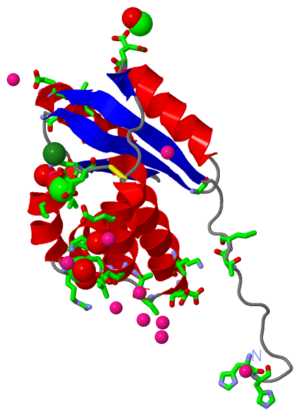
Sites (15, 15)
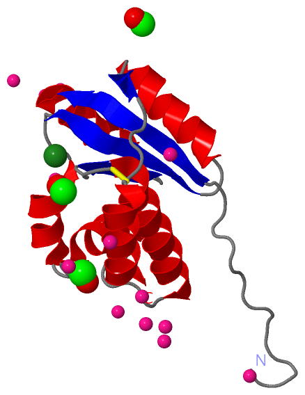
Asymmetric Unit (15, 15)
|
SS Bonds (1, 1)
Asymmetric/Biological Unit
|
||||||||
Cis Peptide Bonds (1, 1)
Asymmetric/Biological Unit
|
||||||||
SAPs(SNPs)/Variants (0, 0)| (no "SAP(SNP)/Variant" information available for 1ZD0) |
PROSITE Motifs (0, 0)| (no "PROSITE Motif" information available for 1ZD0) |
Exons (0, 0)| (no "Exon" information available for 1ZD0) |
Sequences/Alignments
Asymmetric/Biological UnitChain A from PDB Type:PROTEIN Length:141 aligned with Q8U3E5_PYRFU | Q8U3E5 from UniProtKB/TrEMBL Length:142 Alignment length:141 1 | 6 16 26 36 46 56 66 76 86 96 106 116 126 136 Q8U3E5_PYRFU - ----MLEIRTKVGEICISKVWLTDEQINKLFDRFKGDYQVVNAECADKVIFATIIAIKAVKEGRSIAKTVPGEILVRLSGNRQIKEAIKKVGAKEGENYIVTFGENASALLQKILSTLEIKELELERCDLEYAKKAFEDIA 137 SCOP domains -----d1zd0a1 A:9-144 Hypothetical protein PF0523 SCOP domains CATH domains --------------------------------------------------------------------------------------------------------------------------------------------- CATH domains Pfam domains -------------CGI-121-1zd0A01 A:17-144 Pfam domains SAPs(SNPs) --------------------------------------------------------------------------------------------------------------------------------------------- SAPs(SNPs) PROSITE --------------------------------------------------------------------------------------------------------------------------------------------- PROSITE Transcript --------------------------------------------------------------------------------------------------------------------------------------------- Transcript 1zd0 A 4 HHHGSLEIRTKVGEICISKVWLTDEQINKLFDRFKGDYQVVNAECADKVIFATIIAIKAVKEGRSIAKTVPGEILVRLSGNRQIKEAIKKVGAKEGENYIVTFGENASALLQKILSTLEIKELELERCDLEYAKKAFEDIA 144 13 23 33 43 53 63 73 83 93 103 113 123 133 143
|
||||||||||||||||||||
SCOP Domains (1, 1)
Asymmetric/Biological Unit

|
CATH Domains (0, 0)| (no "CATH Domain" information available for 1ZD0) |
Pfam Domains (1, 1)
Asymmetric/Biological Unit
|
Gene Ontology (0, 0)|
Asymmetric/Biological Unit(hide GO term definitions)
(no "Gene Ontology" information available for 1ZD0)
|
Interactive Views
|
|||||||||||||||||||||||||||||||||||||||||||||||||||||||||||||||||||||||||||||||||||||||||||||||||||||||||||||||||||||||||||||||||||||||||||||||||||||||||||||||||||||||||||||||||||||||||||||||||||||||||||||||||||||||||||||||||||||||
Still Images
|
||||||||||||||||
Databases
|
||||||||||||||||||||||||||||||||||||||||||||||||||||||||||||||||||||||||||||||||||||||||||||||||||||||||||||||||||||||||||||||||||||||||||||||||||||||||||||||||
Analysis Tools
|
|||||||||||||||||||||||||||||||||||||||||||||||||||||||||||||
Entries Sharing at Least One Protein Chain (UniProt ID)
Related Entries Specified in the PDB File
|
|