| molecular function |
|---|
| | GO:0019003 | | GDP binding | | Interacting selectively and non-covalently with GDP, guanosine 5'-diphosphate. |
| | GO:0005525 | | GTP binding | | Interacting selectively and non-covalently with GTP, guanosine triphosphate. |
| | GO:0003924 | | GTPase activity | | Catalysis of the reaction: GTP + H2O = GDP + phosphate. |
| | GO:0000166 | | nucleotide binding | | Interacting selectively and non-covalently with a nucleotide, any compound consisting of a nucleoside that is esterified with (ortho)phosphate or an oligophosphate at any hydroxyl group on the ribose or deoxyribose. |
| biological process |
|---|
| | GO:0052405 | | negative regulation by host of symbiont molecular function | | Any process in which an organism stops, prevents, or reduces the frequency, rate or extent of the functional activity of symbiont proteins. The symbiont is defined as the smaller of the organisms involved in a symbiotic interaction. |
| | GO:0045921 | | positive regulation of exocytosis | | Any process that activates or increases the frequency, rate or extent of exocytosis. |
| | GO:0015031 | | protein transport | | The directed movement of proteins into, out of or within a cell, or between cells, by means of some agent such as a transporter or pore. |
| | GO:0032880 | | regulation of protein localization | | Any process that modulates the frequency, rate or extent of any process in which a protein is transported to, or maintained in, a specific location. |
| | GO:0042147 | | retrograde transport, endosome to Golgi | | The directed movement of membrane-bounded vesicles from endosomes back to the trans-Golgi network where they are recycled for further rounds of transport. |
| | GO:0007264 | | small GTPase mediated signal transduction | | Any series of molecular signals in which a small monomeric GTPase relays one or more of the signals. |
| | GO:0006810 | | transport | | The directed movement of substances (such as macromolecules, small molecules, ions) or cellular components (such as complexes and organelles) into, out of or within a cell, or between cells, or within a multicellular organism by means of some agent such as a transporter, pore or motor protein. |
| cellular component |
|---|
| | GO:0005794 | | Golgi apparatus | | A compound membranous cytoplasmic organelle of eukaryotic cells, consisting of flattened, ribosome-free vesicles arranged in a more or less regular stack. The Golgi apparatus differs from the endoplasmic reticulum in often having slightly thicker membranes, appearing in sections as a characteristic shallow semicircle so that the convex side (cis or entry face) abuts the endoplasmic reticulum, secretory vesicles emerging from the concave side (trans or exit face). In vertebrate cells there is usually one such organelle, while in invertebrates and plants, where they are known usually as dictyosomes, there may be several scattered in the cytoplasm. The Golgi apparatus processes proteins produced on the ribosomes of the rough endoplasmic reticulum; such processing includes modification of the core oligosaccharides of glycoproteins, and the sorting and packaging of proteins for transport to a variety of cellular locations. Three different regions of the Golgi are now recognized both in terms of structure and function: cis, in the vicinity of the cis face, trans, in the vicinity of the trans face, and medial, lying between the cis and trans regions. |
| | GO:0000139 | | Golgi membrane | | The lipid bilayer surrounding any of the compartments of the Golgi apparatus. |
| | GO:0031410 | | cytoplasmic vesicle | | A vesicle found in the cytoplasm of a cell. |
| | GO:0005829 | | cytosol | | The part of the cytoplasm that does not contain organelles but which does contain other particulate matter, such as protein complexes. |
| | GO:0005783 | | endoplasmic reticulum | | The irregular network of unit membranes, visible only by electron microscopy, that occurs in the cytoplasm of many eukaryotic cells. The membranes form a complex meshwork of tubular channels, which are often expanded into slitlike cavities called cisternae. The ER takes two forms, rough (or granular), with ribosomes adhering to the outer surface, and smooth (with no ribosomes attached). |
| | GO:0005789 | | endoplasmic reticulum membrane | | The lipid bilayer surrounding the endoplasmic reticulum. |
| | GO:0005768 | | endosome | | A vacuole to which materials ingested by endocytosis are delivered. |
| | GO:0070062 | | extracellular exosome | | A vesicle that is released into the extracellular region by fusion of the limiting endosomal membrane of a multivesicular body with the plasma membrane. Extracellular exosomes, also simply called exosomes, have a diameter of about 40-100 nm. |
| | GO:0005770 | | late endosome | | A prelysosomal endocytic organelle differentiated from early endosomes by lower lumenal pH and different protein composition. Late endosomes are more spherical than early endosomes and are mostly juxtanuclear, being concentrated near the microtubule organizing center. |
| | GO:0005764 | | lysosome | | A small lytic vacuole that has cell cycle-independent morphology and is found in most animal cells and that contains a variety of hydrolases, most of which have their maximal activities in the pH range 5-6. The contained enzymes display latency if properly isolated. About 40 different lysosomal hydrolases are known and lysosomes have a great variety of morphologies and functions. |
| | GO:0016020 | | membrane | | A lipid bilayer along with all the proteins and protein complexes embedded in it an attached to it. |
| | GO:0045335 | | phagocytic vesicle | | A membrane-bounded intracellular vesicle that arises from the ingestion of particulate material by phagocytosis. |
| | GO:0030670 | | phagocytic vesicle membrane | | The lipid bilayer surrounding a phagocytic vesicle. |
| | GO:0005886 | | plasma membrane | | The membrane surrounding a cell that separates the cell from its external environment. It consists of a phospholipid bilayer and associated proteins. |
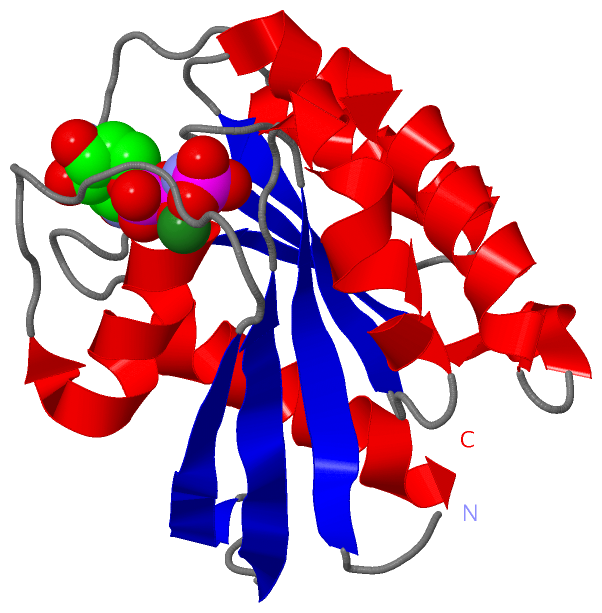
 Description
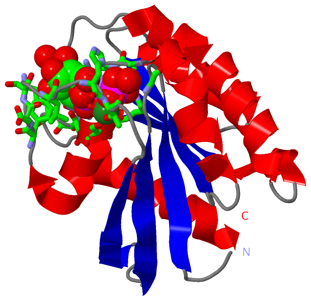