Show PDB file:   
         Plain Text   HTML   (compressed file size)
QuickSearch:   
by PDB,NDB,UniProt,PROSITE Code or Search Term(s)  
(-)Asym./Biol. Unit
(-)Asym./Biol. Unit - sites
collapse expand < >
Image Asym./Biol. Unit
Asym./Biol. Unit  (Jmol Viewer)
Image Asym./Biol. Unit - sites
Asym./Biol. Unit - sites  (Jmol Viewer)

(-) Description

Title :  CRYSTAL STRUCTURE OF UREIDOGLYCOLATE HYDROLASE (ALLA) FROM ESCHERICHIA COLI O157:H7
 
Authors :  S. Raymond, A. Tocilj, A. Matte, M. Cygler, Montreal-Kingston Bacter Structural Genomics Initiative (Bsgi)
Date :  01 Feb 05  (Deposition) - 18 Oct 05  (Release) - 13 Jul 11  (Revision)
Method :  X-RAY DIFFRACTION
Resolution :  1.71
Chains :  Asym./Biol. Unit :  A,B
Keywords :  Alla, Ureidoglycolate, Hydrolase, Purine Metabolism, Structural Genomics, Montreal-Kingston Bacterial Structural Genomics Initiative, Bsgi (Keyword Search: [Gene Ontology, PubMed, Web (Google))
 
Reference :  S. Raymond, A. Tocilj, E. Ajamian, Y. Li, M. -N. Hung, A. Matte, M. Cygle
Crystal Structure Of Ureidoglycolate Hydrolase (Alla) From Escherichia Coli O157:H7
Proteins V. 61 454 2005
PubMed-ID: 16114032  |  Reference-DOI: 10.1002/PROT.20537
(for further references see the PDB file header)

(-) Compounds

Molecule 1 - UREIDOGLYCOLATE HYDROLASE
    ChainsA, B
    EC Number3.5.3.19
    EngineeredYES
    Expression SystemESCHERICHIA COLI
    Expression System PlasmidPET20B
    Expression System Taxid562
    Expression System Vector TypePLASMID
    GeneALLA
    Organism ScientificESCHERICHIA COLI
    Organism Taxid83334

 Structural Features

(-) Chains, Units

  12
Asymmetric/Biological Unit AB

Summary Information (see also Sequences/Alignments below)
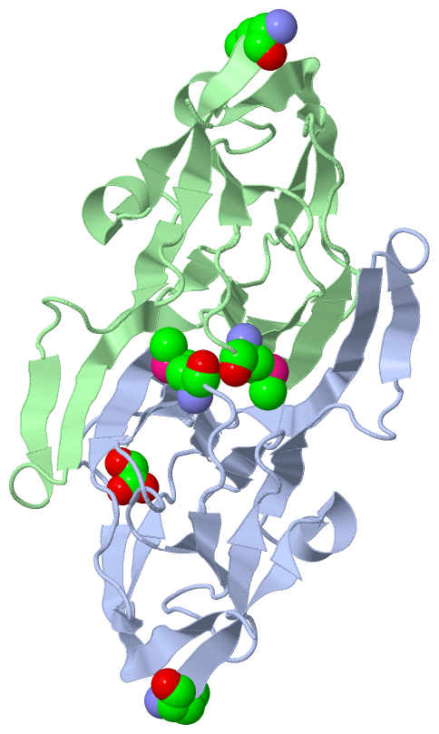
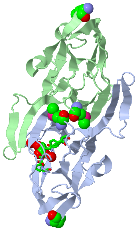
(-) Ligands, Modified Residues, Ions  (2, 5)

Asymmetric/Biological Unit (2, 5)
No.NameCountTypeFull Name
1GLV1Ligand/IonGLYOXYLIC ACID
2MSE4Mod. Amino AcidSELENOMETHIONINE

(-) Sites  (1, 1)

Asymmetric Unit (1, 1)
No.NameEvidenceResiduesDescription
1AC1SOFTWAREGLU A:72 , GLN A:79 , PHE A:81 , PRO A:126 , LEU A:127 , HOH A:1020 , HOH A:1088 , HOH A:1113BINDING SITE FOR RESIDUE GLV A 1001

(-) SS Bonds  (0, 0)

(no "SS Bond" information available for 1YQC)

(-) Cis Peptide Bonds  (2, 2)

Asymmetric/Biological Unit
No.Residues
1Leu A:64 -Pro A:65
2Leu B:64 -Pro B:65

 Sequence-Structure Mapping

(-) SAPs(SNPs)/Variants  (0, 0)

(no "SAP(SNP)/Variant" information available for 1YQC)

(-) PROSITE Motifs  (0, 0)

(no "PROSITE Motif" information available for 1YQC)

(-) Exons   (0, 0)

(no "Exon" information available for 1YQC)

(-) Sequences/Alignments

Asymmetric/Biological Unit
   Reformat: Number of residues per line =  ('0' or empty: single-line sequence representation)
  Number of residues per labelling interval =   
  UniProt sequence: complete  aligned part    
   Show mapping: SCOP domains CATH domains Pfam domains Secondary structure (by author)
SAPs(SNPs) PROSITE motifs Exons
(details for a mapped element are shown in a popup box when the mouse pointer rests over it)
Chain A from PDB  Type:PROTEIN  Length:160
 aligned with ALLA_ECO57 | P63486 from UniProtKB/Swiss-Prot  Length:160

    Alignment length:160
                                    10        20        30        40        50        60        70        80        90       100       110       120       130       140       150       160
           ALLA_ECO57     1 MKLQVLPLSQEAFSAYGDVIETQQRDFFHINNGLVERYHDLALVEILEQDRTLISINRAQPANLPLTIHELERHPLGTQAFIPMKGEVFVVVVALGDDKPDLSTLRAFITNGEQGVNYHRNVWHHPLFAWQRVTDFLTIDRGGSDNCDVESIPEQELCFA 160
               SCOP domains d1yqca1 A:1-160 Ureidoglycolate hydrolase AllA                                                                                                                   SCOP domains
               CATH domains -1yqcA00 A:2-160 Ureidoglycolate hydrolase                                                                                                                       CATH domains
               Pfam domains ---------------------------------------------------------------------------------------------------------------------------------------------------------------- Pfam domains
         Sec.struct. author .eeeeee.hhhhhh..eeee......eeee....eeeeeeeee....hhhh.eeeeeee.......eeeeeee.....eeeee......eeeee...........eeee.....eeee....ee.........eeeeeee.......eeeeeeeeeee.. Sec.struct. author
                 SAPs(SNPs) ---------------------------------------------------------------------------------------------------------------------------------------------------------------- SAPs(SNPs)
                    PROSITE ---------------------------------------------------------------------------------------------------------------------------------------------------------------- PROSITE
                 Transcript ---------------------------------------------------------------------------------------------------------------------------------------------------------------- Transcript
                 1yqc A   1 mKLQVLPLSQEAFSAYGDVIETQQRDFFHINNGLVERYHDLALVEILEQDRTLISINRAQPANLPLTIHELERHPLGTQAFIPmKGEVFVVVVALGDDKPDLSTLRAFITNGEQGVNYHRNVWHHPLFAWQRVTDFLTIDRGGSDNCDVESIPEQELCFA 160
                            |       10        20        30        40        50        60        70        80   |    90       100       110       120       130       140       150       160
                            |                                                                                 84-MSE                                                                        
                            1-MSE                                                                                                                                                           

Chain B from PDB  Type:PROTEIN  Length:160
 aligned with ALLA_ECO57 | P63486 from UniProtKB/Swiss-Prot  Length:160

    Alignment length:160
                                    10        20        30        40        50        60        70        80        90       100       110       120       130       140       150       160
           ALLA_ECO57     1 MKLQVLPLSQEAFSAYGDVIETQQRDFFHINNGLVERYHDLALVEILEQDRTLISINRAQPANLPLTIHELERHPLGTQAFIPMKGEVFVVVVALGDDKPDLSTLRAFITNGEQGVNYHRNVWHHPLFAWQRVTDFLTIDRGGSDNCDVESIPEQELCFA 160
               SCOP domains d1yqcb_ B: Ureidoglycolate hydrolase AllA                                                                                                                        SCOP domains
               CATH domains -1yqcB00 B:2-160 Ureidoglycolate hydrolase                                                                                                                       CATH domains
           Pfam domains (1) Ureidogly_hydro-1yqcB01 B:1-158                                                                                                                               -- Pfam domains (1)
           Pfam domains (2) Ureidogly_hydro-1yqcB02 B:1-158                                                                                                                               -- Pfam domains (2)
         Sec.struct. author .eeeeee.hhhhhh..eeee......eeee....eeeeeeeee.........eeeeeee.......eeeeeee.....eeeee......eeeee......hhhhheeee.....eeee....ee.........eeeeeee.......eeeeeeeeeee.. Sec.struct. author
                 SAPs(SNPs) ---------------------------------------------------------------------------------------------------------------------------------------------------------------- SAPs(SNPs)
                    PROSITE ---------------------------------------------------------------------------------------------------------------------------------------------------------------- PROSITE
                 Transcript ---------------------------------------------------------------------------------------------------------------------------------------------------------------- Transcript
                 1yqc B   1 mKLQVLPLSQEAFSAYGDVIETQQRDFFHINNGLVERYHDLALVEILEQDRTLISINRAQPANLPLTIHELERHPLGTQAFIPmKGEVFVVVVALGDDKPDLSTLRAFITNGEQGVNYHRNVWHHPLFAWQRVTDFLTIDRGGSDNCDVESIPEQELCFA 160
                            |       10        20        30        40        50        60        70        80   |    90       100       110       120       130       140       150       160
                            1-MSE                                                                             84-MSE                                                                        

   Legend:   → Mismatch (orange background)
  - → Gap (green background, '-', border residues have a numbering label)
    → Modified Residue (blue background, lower-case, 'x' indicates undefined single-letter code, labelled with number + name)
  x → Chemical Group (purple background, 'x', labelled with number + name, e.g. ACE or NH2)
  extra numbering lines below/above indicate numbering irregularities and modified residue names etc., number ends below/above '|'

 Classification and Annotation

(-) SCOP Domains  (1, 2)

Asymmetric/Biological Unit

(-) CATH Domains  (1, 2)

Asymmetric/Biological Unit
(-)
Class: Mainly Beta (13760)

(-) Pfam Domains  (1, 2)

Asymmetric/Biological Unit

(-) Gene Ontology  (6, 6)

Asymmetric/Biological Unit(hide GO term definitions)
Chain A,B   (ALLA_ECO57 | P63486)
molecular function
    GO:0016829    lyase activity    Catalysis of the cleavage of C-C, C-O, C-N and other bonds by other means than by hydrolysis or oxidation, or conversely adding a group to a double bond. They differ from other enzymes in that two substrates are involved in one reaction direction, but only one in the other direction. When acting on the single substrate, a molecule is eliminated and this generates either a new double bond or a new ring.
    GO:0004848    ureidoglycolate hydrolase activity    Catalysis of the reaction: (S)-ureidoglycolate + H(2)O + 2 H(+) = CO(2) + glyoxylate + 2 NH(4)(+).
    GO:0050385    ureidoglycolate lyase activity    Catalysis of the reaction: (S)-ureidoglycolate = glyoxylate + urea.
biological process
    GO:0000256    allantoin catabolic process    The chemical reactions and pathways resulting in the breakdown of allantoin, (2,5-dioxo-4-imidazolidinyl)urea.
    GO:0006145    purine nucleobase catabolic process    The chemical reactions and pathways resulting in the breakdown of purine nucleobases, one of the two classes of nitrogen-containing ring compounds found in DNA and RNA, which include adenine and guanine.
    GO:0006144    purine nucleobase metabolic process    The chemical reactions and pathways involving purine nucleobases, one of the two classes of nitrogen-containing ring compounds found in DNA and RNA, which include adenine and guanine.

 Visualization

(-) Interactive Views

Asymmetric/Biological Unit
  Complete Structure
    Jena3D(integrated viewing of ligand, site, SAP, PROSITE, SCOP information)
    WebMol | AstexViewer[tm]@PDBe
(Java Applets, require no local installation except for Java; loading may be slow)
    STRAP
(Java WebStart application, automatic local installation, requires Java; full application with system access!)
    RasMol
(require local installation)
    Molscript (VRML)
(requires installation of a VRML viewer; select preferred view via VRML and generate a mono or stereo PDF format file)
 
  Ligands, Modified Residues, Ions
    GLV  [ RasMol | Jena3D ]  +environment [ RasMol | Jena3D ]
    MSE  [ RasMol | Jena3D ]  +environment [ RasMol | Jena3D ]
 
  Sites
    AC1  [ RasMol ]  +environment [ RasMol ]
 
  Cis Peptide Bonds
    Leu A:64 - Pro A:65   [ RasMol ]  
    Leu B:64 - Pro B:65   [ RasMol ]  
 

(-) Still Images

Jmol
  protein: cartoon or spacefill or dots and stick; nucleic acid: cartoon and stick; ligands: spacefill; active site: stick
Molscript
  protein, nucleic acid: cartoon; ligands: spacefill; active site: ball and stick

 Databases and Analysis Tools

(-) Databases

Access by PDB/NDB ID
  1yqc
    Family and Domain InformationProDom | SYSTERS
    General Structural InformationGlycoscienceDB | MMDB | NDB | OCA | PDB | PDBe | PDBj | PDBsum | PDBWiki | PQS | PROTEOPEDIA
    Orientation in MembranesOPM
    Protein SurfaceSURFACE
    Secondary StructureDSSP (structure derived) | HSSP (homology derived)
    Structural GenomicsGeneCensus
    Structural NeighboursCE | VAST
    Structure ClassificationCATH | Dali | SCOP
    Validation and Original DataBMRB Data View | BMRB Restraints Grid | EDS | PROCHECK | RECOORD | WHAT_CHECK
 
Access by UniProt ID/Accession number
  ALLA_ECO57 | P63486
    Comparative Protein Structure ModelsModBase
    Genomic InformationEnsembl
    Protein-protein InteractionDIP
    Sequence, Family and Domain InformationInterPro | Pfam | SMART | UniProtKB/SwissProt
 
Access by Enzyme Classificator   (EC Number)
  3.5.3.19
    General Enzyme InformationBRENDA | EC-PDB | Enzyme | IntEnz
    PathwayKEGG | MetaCyc
 
Access by Disease Identifier   (MIM ID)
  (no 'MIM ID' available)
    Disease InformationOMIM
 
Access by GenAge ID
  (no 'GenAge ID' available)
    Age Related InformationGenAge

(-) Analysis Tools

Access by PDB/NDB ID
    Domain InformationXDom
    Interatomic Contacts of Structural UnitsCSU
    Ligand-protein ContactsLPC
    Protein CavitiescastP
    Sequence and Secondary StructurePDBCartoon
    Structure AlignmentSTRAP(Java WebStart application, automatic local installation, requires Java; full application with system access!)
    Structure and Sequence BrowserSTING
 
Access by UniProt ID/Accession number
  ALLA_ECO57 | P63486
    Protein Disorder PredictionDisEMBL | FoldIndex | GLOBPLOT (for more information see DisProt)

 Related Entries

(-) Entries Sharing at Least One Protein Chain (UniProt ID)

(no "Entries Sharing at Least One Protein Chain" available for 1YQC)

(-) Related Entries Specified in the PDB File

(no "Related Entries Specified in the PDB File" available for 1YQC)