| molecular function |
|---|
| | GO:0005524 | | ATP binding | | Interacting selectively and non-covalently with ATP, adenosine 5'-triphosphate, a universally important coenzyme and enzyme regulator. |
| | GO:0046404 | | ATP-dependent polydeoxyribonucleotide 5'-hydroxyl-kinase activity | | Catalysis of the reaction: ATP + 5'-dephospho-DNA = ADP + 5'-phospho-DNA. |
| | GO:0003824 | | catalytic activity | | Catalysis of a biochemical reaction at physiological temperatures. In biologically catalyzed reactions, the reactants are known as substrates, and the catalysts are naturally occurring macromolecular substances known as enzymes. Enzymes possess specific binding sites for substrates, and are usually composed wholly or largely of protein, but RNA that has catalytic activity (ribozyme) is often also regarded as enzymatic. |
| | GO:0003690 | | double-stranded DNA binding | | Interacting selectively and non-covalently with double-stranded DNA. |
| | GO:0016787 | | hydrolase activity | | Catalysis of the hydrolysis of various bonds, e.g. C-O, C-N, C-C, phosphoric anhydride bonds, etc. Hydrolase is the systematic name for any enzyme of EC class 3. |
| | GO:0016301 | | kinase activity | | Catalysis of the transfer of a phosphate group, usually from ATP, to a substrate molecule. |
| | GO:0000166 | | nucleotide binding | | Interacting selectively and non-covalently with a nucleotide, any compound consisting of a nucleoside that is esterified with (ortho)phosphate or an oligophosphate at any hydroxyl group on the ribose or deoxyribose. |
| | GO:0019201 | | nucleotide kinase activity | | Catalysis of the reaction: ATP + nucleoside monophosphate = ADP + nucleoside diphosphate. |
| | GO:0046403 | | polynucleotide 3'-phosphatase activity | | Catalysis of the reaction: 3'-phosphopolynucleotide + H2O = a polynucleotide + phosphate. Hydrolyzes the free 3'-phosphate resulting from single strand breaks in DNA due to oxidative damage. |
| | GO:0016740 | | transferase activity | | Catalysis of the transfer of a group, e.g. a methyl group, glycosyl group, acyl group, phosphorus-containing, or other groups, from one compound (generally regarded as the donor) to another compound (generally regarded as the acceptor). Transferase is the systematic name for any enzyme of EC class 2. |
| biological process |
|---|
| | GO:0098504 | | DNA 3' dephosphorylation involved in DNA repair | | Any 3' DNA dephosphorylation that is involved in the process of DNA repair. |
| | GO:0042769 | | DNA damage response, detection of DNA damage | | The series of events required to receive a stimulus indicating DNA damage has occurred and convert it to a molecular signal. |
| | GO:0006281 | | DNA repair | | The process of restoring DNA after damage. Genomes are subject to damage by chemical and physical agents in the environment (e.g. UV and ionizing radiations, chemical mutagens, fungal and bacterial toxins, etc.) and by free radicals or alkylating agents endogenously generated in metabolism. DNA is also damaged because of errors during its replication. A variety of different DNA repair pathways have been reported that include direct reversal, base excision repair, nucleotide excision repair, photoreactivation, bypass, double-strand break repair pathway, and mismatch repair pathway. |
| | GO:0006974 | | cellular response to DNA damage stimulus | | Any process that results in a change in state or activity of a cell (in terms of movement, secretion, enzyme production, gene expression, etc.) as a result of a stimulus indicating damage to its DNA from environmental insults or errors during metabolism. |
| | GO:0016311 | | dephosphorylation | | The process of removing one or more phosphoric (ester or anhydride) residues from a molecule. |
| | GO:0008152 | | metabolic process | | The chemical reactions and pathways, including anabolism and catabolism, by which living organisms transform chemical substances. Metabolic processes typically transform small molecules, but also include macromolecular processes such as DNA repair and replication, and protein synthesis and degradation. |
| | GO:0046939 | | nucleotide phosphorylation | | The process of introducing one or more phosphate groups into a nucleotide to produce a phosphorylated nucleoside. |
| | GO:0016310 | | phosphorylation | | The process of introducing a phosphate group into a molecule, usually with the formation of a phosphoric ester, a phosphoric anhydride or a phosphoric amide. |
| | GO:0098506 | | polynucleotide 3' dephosphorylation | | The process of removing one or more phosphate groups from the 3' end of a polynucleotide. |
| | GO:0051973 | | positive regulation of telomerase activity | | Any process that activates or increases the frequency, rate or extent of telomerase activity, the catalysis of the reaction: deoxynucleoside triphosphate + DNA(n) = diphosphate + DNA(n+1). |
| | GO:1904355 | | positive regulation of telomere capping | | Any process that activates or increases the frequency, rate or extent of telomere capping. |
| | GO:0032212 | | positive regulation of telomere maintenance via telomerase | | Any process that activates or increases the frequency, rate or extent of the addition of telomeric repeats by telomerase. |
| | GO:0006979 | | response to oxidative stress | | Any process that results in a change in state or activity of a cell or an organism (in terms of movement, secretion, enzyme production, gene expression, etc.) as a result of oxidative stress, a state often resulting from exposure to high levels of reactive oxygen species, e.g. superoxide anions, hydrogen peroxide (H2O2), and hydroxyl radicals. |
| cellular component |
|---|
| | GO:0016020 | | membrane | | A lipid bilayer along with all the proteins and protein complexes embedded in it an attached to it. |
| | GO:0005730 | | nucleolus | | A small, dense body one or more of which are present in the nucleus of eukaryotic cells. It is rich in RNA and protein, is not bounded by a limiting membrane, and is not seen during mitosis. Its prime function is the transcription of the nucleolar DNA into 45S ribosomal-precursor RNA, the processing of this RNA into 5.8S, 18S, and 28S components of ribosomal RNA, and the association of these components with 5S RNA and proteins synthesized outside the nucleolus. This association results in the formation of ribonucleoprotein precursors; these pass into the cytoplasm and mature into the 40S and 60S subunits of the ribosome. |
| | GO:0005634 | | nucleus | | A membrane-bounded organelle of eukaryotic cells in which chromosomes are housed and replicated. In most cells, the nucleus contains all of the cell's chromosomes except the organellar chromosomes, and is the site of RNA synthesis and processing. In some species, or in specialized cell types, RNA metabolism or DNA replication may be absent. |
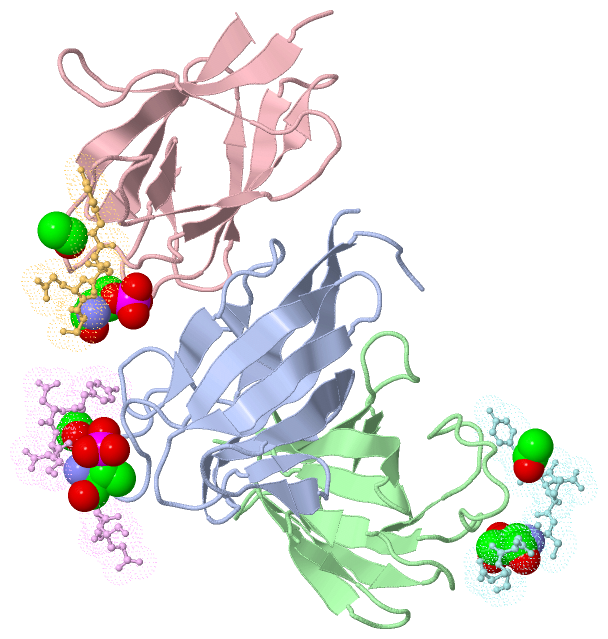
 Description
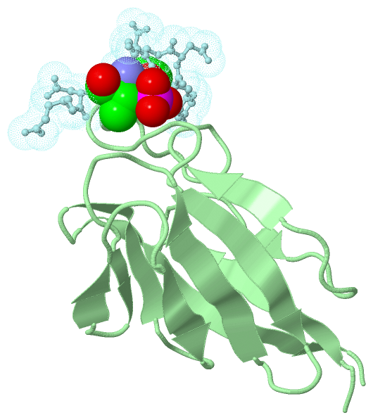