|
|
|
|
 Description
|
|
Compounds
|
||||||||||||||||||||||||||||||||||||||||||||||||||||
Chains, Units
Summary Information (see also Sequences/Alignments below) |
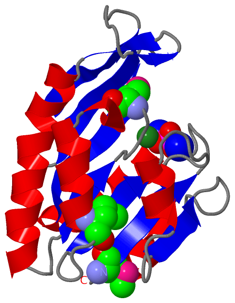
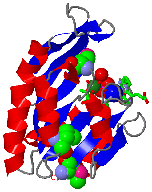
Ligands, Modified Residues, Ions (4, 7)| Asymmetric/Biological Unit (4, 7) |
Sites (4, 4)
Asymmetric Unit (4, 4)
|
SS Bonds (0, 0)| (no "SS Bond" information available for 1YE8) |
Cis Peptide Bonds (1, 1)
Asymmetric/Biological Unit
|
||||||||
SAPs(SNPs)/Variants (0, 0)| (no "SAP(SNP)/Variant" information available for 1YE8) |
PROSITE Motifs (0, 0)| (no "PROSITE Motif" information available for 1YE8) |
Exons (0, 0)| (no "Exon" information available for 1YE8) |
Sequences/Alignments
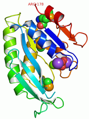
Asymmetric/Biological UnitChain A from PDB Type:PROTEIN Length:172 aligned with NTPTH_AQUAE | O67322 from UniProtKB/Swiss-Prot Length:178 Alignment length:178 10 20 30 40 50 60 70 80 90 100 110 120 130 140 150 160 170 NTPTH_AQUAE 1 MKIIITGEPGVGKTTLVKKIVERLGKRAIGFWTEEVRDPETKKRTGFRIITTEGKKKIFSSKFFTSKKLVGSYGVNVQYFEELAIPILERAYREAKKDRRKVIIIDEIGKMELFSKKFRDLVRQIMHDPNVNVVATIPIRDVHPLVKEIRRLPGAVLIELTPENRDVILEDILSLLER 178 SCOP domains d1ye8a1 A:1-178 Hypothetical kinase-l ike protein Aq_1292 SCOP domains CATH domains ---------------------------------------------------------------------------------------------------------------------------------------------------------------------------------- CATH domains Pfam domains -NTPase_1-1ye8A01 A:2-173 ----- Pfam domains SAPs(SNPs) ---------------------------------------------------------------------------------------------------------------------------------------------------------------------------------- SAPs(SNPs) PROSITE ---------------------------------------------------------------------------------------------------------------------------------------------------------------------------------- PROSITE Transcript ---------------------------------------------------------------------------------------------------------------------------------------------------------------------------------- Transcript 1ye8 A 1 mKIIITGEPGVGKTTLVKKIVERLGKRAIGFWTEEVR------RTGFRIITTEGKKKIFSSKFFTSKKLVGSYGVNVQYFEELAIPILERAYREAKKDRRKVIIIDEIGKmELFSKKFRDLVRQImHDPNVNVVATIPIRDVHPLVKEIRRLPGAVLIELTPENRDVILEDILSLLER 178 | 10 20 30 | - | 50 60 70 80 90 100 110| 120 | 130 140 150 160 170 | 37 44 111-MSE 126-MSE 1-MSE
|
||||||||||||||||||||
SCOP Domains (1, 1)
Asymmetric/Biological Unit
 |
CATH Domains (0, 0)| (no "CATH Domain" information available for 1YE8) |
Pfam Domains (1, 1)
Asymmetric/Biological Unit
|
Gene Ontology (6, 6)|
Asymmetric/Biological Unit(hide GO term definitions) Chain A (NTPTH_AQUAE | O67322)
|
||||||||||||||||||||||||||||||||||||||||||||||||
Interactive Views
|
|||||||||||||||||||||||||||||||||||||||||||||||||||||||||||||||||||||||||||||||||||||||||||||||||||||||||||||||||||||||||||||||||||||||||||||||||||||||||||||||||
Still Images
|
||||||||||||||||
Databases
|
||||||||||||||||||||||||||||||||||||||||||||||||||||||||||||||||||||||||||||||||||||||||||||||||||||||||||||||||||||||||||||||||||||||||||||||||||||||||||||||||
Analysis Tools
|
|||||||||||||||||||||||||||||||||||||||||||||||||||||||||||||
Entries Sharing at Least One Protein Chain (UniProt ID)
Related Entries Specified in the PDB File
|
|