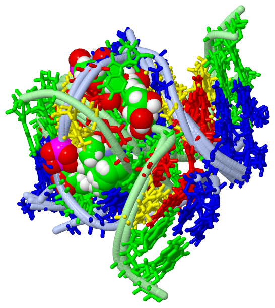
Show PDB file:   
         Plain Text   HTML   (compressed file size)
QuickSearch:   
by PDB,NDB,UniProt,PROSITE Code or Search Term(s)  
(-)NMR Structure - manually
(-)NMR Structure - model 1
(-)NMR Structure - model 1, sites
(-)NMR Structure - all models
collapse expand < >
Image NMR Structure - manually
NMR Structure - manually  (Jmol Viewer)
Image NMR Structure - model 1
NMR Structure - model 1  (Jmol Viewer)
Image NMR Structure - model 1, sites
NMR Structure - model 1, sites  (Jmol Viewer)
Image NMR Structure - all models
NMR Structure - all models  (Jmol Viewer)

(-) Description

Title :  METHYLATION OF CYTOSINE AT C5 IN A CPG SEQUENCE CONTEXT CAUSES A CONFORMATIONAL SWITCH OF A BENZO[A]PYRENE DIOL EPOXIDE-N2-GUANINE ADDUCT IN DNA FROM A MINOR GROOVE ALIGNMENT TO INTERCALATION WITH BASE DISPLACEMENT
 
Authors :  N. Zhang, C. Lin, X. Huang, A. Kolbanovskiy, B. E. Hingerty, S. Amin, S. Broyde, N. E. Geacintov, D. J. Patel
Date :  15 Dec 04  (Deposition) - 22 Mar 05  (Release) - 24 Feb 09  (Revision)
Method :  SOLUTION NMR
Resolution :  NOT APPLICABLE
Chains :  NMR Structure  :  A,B  (9x)
Keywords :  Conformational Switch, Cytosine Methylation, P53 Mutation Hot Spot, Dna Adduct, Benzo[A]Pyrene, Bpde (Keyword Search: [Gene Ontology, PubMed, Web (Google))
 
Reference :  N. Zhang, C. Lin, X. Huang, A. Kolbanovskiy, B. E. Hingerty, S. Amin, S. Broyde, N. E. Geacintov, D. J. Patel
Methylation Of Cytosine At C5 In A Cpg Sequence Context Causes A Conformational Switch Of A Benzo[A]Pyrene Diol Epoxide-N2-Guanine Adduct In Dna From A Minor Groove Alignment To Intercalation With Base Displacement.
J. Mol. Biol. V. 346 951 2005
PubMed-ID: 15701509  |  Reference-DOI: 10.1016/J.JMB.2004.12.027
(for further references see the PDB file header)

(-) Compounds

Molecule 1 - 5'-D(*CP*CP*AP*TP*(5CM)P*(BPG)P*CP*TP*AP*CP*C)- 3'
    ChainsA
    EngineeredYES
    Other DetailsBP ATTACHED TO G6, METHYLATION OF C5
    SyntheticYES
 
Molecule 2 - 5'-D(*GP*GP*TP*AP*GP*CP*GP*AP*TP*GP*G)-3'
    ChainsB
    EngineeredYES
    SyntheticYES

 Structural Features

(-) Chains, Units

  
NMR Structure (9x)

Summary Information (see also Sequences/Alignments below)

(-) Ligands, Modified Residues, Ions  (2, 2)

NMR Structure (2, 2)
No.NameCountTypeFull Name
15CM1Mod. Nucleotide5-METHYL-2'-DEOXY-CYTIDINE-5'-MONOPHOSPHATE
2BAP1Ligand/Ion1,2,3-TRIHYDROXY-1,2,3,4-TETRAHYDROBENZO[A]PYRENE

(-) Sites  (1, 1)

NMR Structure (1, 1)
No.NameEvidenceResiduesDescription
1AC1SOFTWARE5CM A:5 , DG A:6 , DC A:7 , DT A:8 , DG B:16 , DG B:18BINDING SITE FOR RESIDUE BAP A 23

(-) SS Bonds  (0, 0)

(no "SS Bond" information available for 1Y9H)

(-) Cis Peptide Bonds  (0, 0)

(no "Cis Peptide Bond" information available for 1Y9H)

 Sequence-Structure Mapping

(-) SAPs(SNPs)/Variants  (0, 0)

(no "SAP(SNP)/Variant" information available for 1Y9H)

(-) PROSITE Motifs  (0, 0)

(no "PROSITE Motif" information available for 1Y9H)

(-) Exons   (0, 0)

(no "Exon" information available for 1Y9H)

(-) Sequences/Alignments

NMR Structure
   Reformat: Number of residues per line =  ('0' or empty: single-line sequence representation)
  Number of residues per labelling interval =   
  UniProt sequence: complete  aligned part    
   Show mapping: SCOP domains CATH domains Pfam domains Secondary structure (by author)
SAPs(SNPs) PROSITE motifs Exons
(details for a mapped element are shown in a popup box when the mouse pointer rests over it)
Chain A from PDB  Type:DNA  Length:11
                                          
                  1y9h A  1 CCATcGCTACC 11
                                |   10 
                                |      
                                5-5CM  

Chain B from PDB  Type:DNA  Length:11
                                          
                  1y9h B 12 GGTAGCGATGG 22
                                    21 

   Legend:   → Mismatch (orange background)
  - → Gap (green background, '-', border residues have a numbering label)
    → Modified Residue (blue background, lower-case, 'x' indicates undefined single-letter code, labelled with number + name)
  x → Chemical Group (purple background, 'x', labelled with number + name, e.g. ACE or NH2)
  extra numbering lines below/above indicate numbering irregularities and modified residue names etc., number ends below/above '|'

 Classification and Annotation

(-) SCOP Domains  (0, 0)

(no "SCOP Domain" information available for 1Y9H)

(-) CATH Domains  (0, 0)

(no "CATH Domain" information available for 1Y9H)

(-) Pfam Domains  (0, 0)

(no "Pfam Domain" information available for 1Y9H)

(-) Gene Ontology  (0, 0)

NMR Structure(hide GO term definitions)
    (no "Gene Ontology" information available for 1Y9H)

 Visualization

(-) Interactive Views

NMR Structure
  Complete Structure
    Jena3D(integrated viewing of ligand, site, SAP, PROSITE, SCOP information)
    WebMol | AstexViewer[tm]@PDBe
(Java Applets, require no local installation except for Java; loading may be slow)
    STRAP
(Java WebStart application, automatic local installation, requires Java; full application with system access!)
    RasMol
(require local installation)
    Molscript (VRML)
(requires installation of a VRML viewer; select preferred view via VRML and generate a mono or stereo PDF format file)
 
  Ligands, Modified Residues, Ions
    5CM  [ RasMol | Jena3D ]  +environment [ RasMol | Jena3D ]
    BAP  [ RasMol | Jena3D ]  +environment [ RasMol | Jena3D ]
 
  Sites
    AC1  [ RasMol ]  +environment [ RasMol ]
 
  Cis Peptide Bonds
(no "Cis Peptide Bonds" information available for 1y9h)
 

(-) Still Images

Jmol
  protein: cartoon or spacefill or dots and stick; nucleic acid: cartoon and stick; ligands: spacefill; active site: stick
Molscript
  protein, nucleic acid: cartoon; ligands: spacefill; active site: ball and stick

 Databases and Analysis Tools

(-) Databases

Access by PDB/NDB ID
  1y9h
    Family and Domain InformationProDom | SYSTERS
    General Structural InformationGlycoscienceDB | MMDB | NDB | OCA | PDB | PDBe | PDBj | PDBsum | PDBWiki | PQS | PROTEOPEDIA
    Orientation in MembranesOPM
    Protein SurfaceSURFACE
    Secondary StructureDSSP | HSSP
    Structural GenomicsGeneCensus
    Structural NeighboursCE | VAST
    Structure ClassificationCATH | Dali | SCOP
    Validation and Original DataBMRB Data View | BMRB Restraints Grid | EDS | PROCHECK | RECOORD | WHAT_CHECK
 
Access by UniProt ID/Accession number
  (no 'UniProt ID/Accession number' available) |
    Comparative Protein Structure ModelsModBase
    Genomic InformationEnsembl
    Protein-protein InteractionDIP
    Sequence, Family and Domain InformationInterPro | Pfam | SMART | UniProt
 
Access by Enzyme Classificator   (EC Number)
  (no 'Enzyme Classificator' available)
    General Enzyme InformationBRENDA | EC-PDB | Enzyme | IntEnz
    PathwayKEGG | MetaCyc
 
Access by Disease Identifier   (MIM ID)
  (no 'MIM ID' available)
    Disease InformationOMIM
 
Access by GenAge ID
  (no 'GenAge ID' available)
    Age Related InformationGenAge

(-) Analysis Tools

Access by PDB/NDB ID
    Domain InformationXDom
    Interatomic Contacts of Structural UnitsCSU
    Ligand-protein ContactsLPC
    Protein CavitiescastP
    Sequence and Secondary StructurePDBCartoon
    Structure AlignmentSTRAP(Java WebStart application, automatic local installation, requires Java; full application with system access!)
    Structure and Sequence BrowserSTING
 
Access by UniProt ID/Accession number
  (no 'UniProt ID/Accession number' available) |
    Protein Disorder PredictionDisEMBL | FoldIndex | GLOBPLOT

 Related Entries

(-) Entries Sharing at Least One Protein Chain (UniProt ID)

(no "Entries Sharing at Least One Protein Chain" available for 1Y9H)

(-) Related Entries Specified in the PDB File

(no "Related Entries Specified in the PDB File" available for 1Y9H)