| molecular function |
|---|
| | GO:0004563 | | beta-N-acetylhexosaminidase activity | | Catalysis of the hydrolysis of terminal non-reducing N-acetyl-D-hexosamine residues in N-acetyl-beta-D-hexosaminides. |
| | GO:0016787 | | hydrolase activity | | Catalysis of the hydrolysis of various bonds, e.g. C-O, C-N, C-C, phosphoric anhydride bonds, etc. Hydrolase is the systematic name for any enzyme of EC class 3. |
| | GO:0016798 | | hydrolase activity, acting on glycosyl bonds | | Catalysis of the hydrolysis of any glycosyl bond. |
| | GO:0004553 | | hydrolase activity, hydrolyzing O-glycosyl compounds | | Catalysis of the hydrolysis of any O-glycosyl bond. |
| biological process |
|---|
| | GO:0005975 | | carbohydrate metabolic process | | The chemical reactions and pathways involving carbohydrates, any of a group of organic compounds based of the general formula Cx(H2O)y. Includes the formation of carbohydrate derivatives by the addition of a carbohydrate residue to another molecule. |
| | GO:0007049 | | cell cycle | | The progression of biochemical and morphological phases and events that occur in a cell during successive cell replication or nuclear replication events. Canonically, the cell cycle comprises the replication and segregation of genetic material followed by the division of the cell, but in endocycles or syncytial cells nuclear replication or nuclear division may not be followed by cell division. |
| | GO:0051301 | | cell division | | The process resulting in division and partitioning of components of a cell to form more cells; may or may not be accompanied by the physical separation of a cell into distinct, individually membrane-bounded daughter cells. |
| | GO:0071555 | | cell wall organization | | A process that results in the assembly, arrangement of constituent parts, or disassembly of the cell wall, the rigid or semi-rigid envelope lying outside the cell membrane of plant, fungal and most prokaryotic cells, maintaining their shape and protecting them from osmotic lysis. |
| | GO:0008152 | | metabolic process | | The chemical reactions and pathways, including anabolism and catabolism, by which living organisms transform chemical substances. Metabolic processes typically transform small molecules, but also include macromolecular processes such as DNA repair and replication, and protein synthesis and degradation. |
| | GO:0009252 | | peptidoglycan biosynthetic process | | The chemical reactions and pathways resulting in the formation of peptidoglycans, any of a class of glycoconjugates found in bacterial cell walls. |
| | GO:0009254 | | peptidoglycan turnover | | The continual breakdown and regeneration of peptidoglycan required to maintain the cell wall. |
| | GO:0009273 | | peptidoglycan-based cell wall biogenesis | | The chemical reactions and pathways resulting in the formation of the peptidoglycan-based cell wall. An example of this process is found in Escherichia coli. |
| | GO:0008360 | | regulation of cell shape | | Any process that modulates the surface configuration of a cell. |
| | GO:0046677 | | response to antibiotic | | Any process that results in a change in state or activity of a cell or an organism (in terms of movement, secretion, enzyme production, gene expression, etc.) as a result of an antibiotic stimulus. An antibiotic is a chemical substance produced by a microorganism which has the capacity to inhibit the growth of or to kill other microorganisms. |
| cellular component |
|---|
| | GO:0005737 | | cytoplasm | | All of the contents of a cell excluding the plasma membrane and nucleus, but including other subcellular structures. |
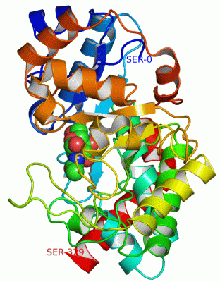
 Description
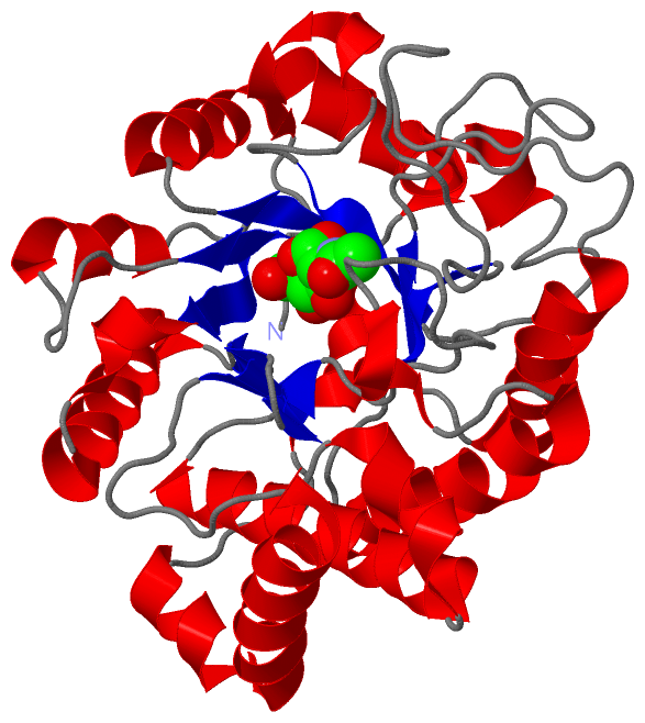
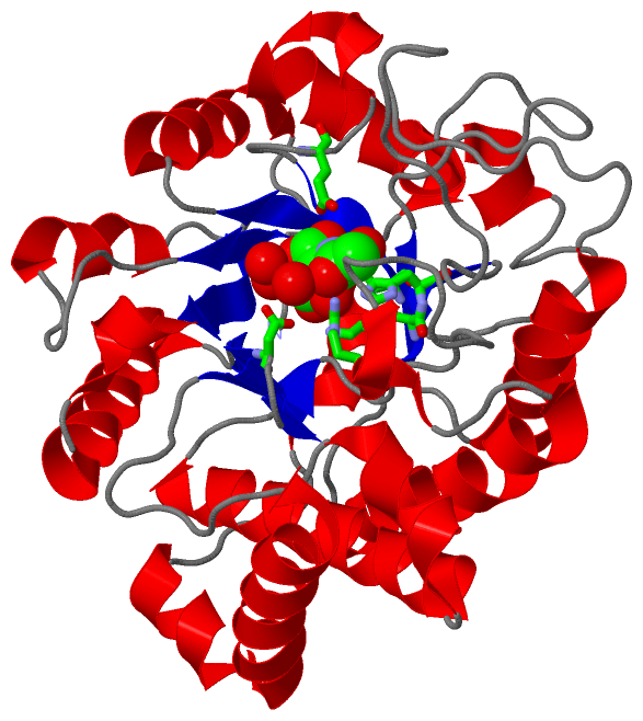