|
|
|
|
 Description
|
|
Compounds
|
||||||||||||||||||||||||||||||||||||||||||||||||
Chains, Units
Summary Information (see also Sequences/Alignments below) |
Ligands, Modified Residues, Ions (4, 6)| Asymmetric/Biological Unit (4, 6) |
Sites (6, 6)
Asymmetric Unit (6, 6)
|
SS Bonds (0, 0)| (no "SS Bond" information available for 1XVI) |
Cis Peptide Bonds (2, 2)
Asymmetric/Biological Unit
|
||||||||||||
SAPs(SNPs)/Variants (0, 0)| (no "SAP(SNP)/Variant" information available for 1XVI) |
PROSITE Motifs (0, 0)| (no "PROSITE Motif" information available for 1XVI) |
Exons (0, 0)| (no "Exon" information available for 1XVI) |
Sequences/Alignments
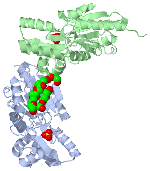
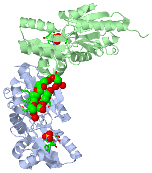
Asymmetric/Biological UnitChain A from PDB Type:PROTEIN Length:232 aligned with MPGP_ECOLI | P76329 from UniProtKB/Swiss-Prot Length:271 Alignment length:232 13 23 33 43 53 63 73 83 93 103 113 123 133 143 153 163 173 183 193 203 213 223 233 MPGP_ECOLI 4 IQQPLLVFSDLDGTLLDSHSYDWQPAAPWLTRLREANVPVILCSSKTSAEMLYLQKTLGLQGLPLIAENGAVIQLAEQWQEIDGFPRIISGISHGEISLVLNTLREKEHFKFTTFDDVDDATIAEWTGLSRSQAALTQLHEASVTLIWRDSDERMAQFTARLNELGLQFMQGARFWHVLDASAGKDQAANWIIATYQQLSGKRPTTLGLGDGPNDAPLLEVMDYAVIVKGLN 235 SCOP domains d1xvia_ A: Putative mannosyl-3-phosphoglycerate phosphatase MPGP (YedP) SCOP domains CATH domains 1xviA01 A:4-92,A:187-235 [code=3.40.50.1000, no name defined] 1xviA02 A:93-186 Putative mannosyl-3-phosphoglycerate phosphatase; domain 2 1xviA01 A:4-92,A:187-235 CATH domains Pfam domains ---------------------------------------------------------------------------------------------------------------------------------------------------------------------------------------------------------------------------------------- Pfam domains SAPs(SNPs) ---------------------------------------------------------------------------------------------------------------------------------------------------------------------------------------------------------------------------------------- SAPs(SNPs) PROSITE ---------------------------------------------------------------------------------------------------------------------------------------------------------------------------------------------------------------------------------------- PROSITE Transcript ---------------------------------------------------------------------------------------------------------------------------------------------------------------------------------------------------------------------------------------- Transcript 1xvi A 4 IQQPLLVFSDLDGTLLDSHSYDWQPAAPWLTRLREANVPVILCSSKTSAEMLYLQKTLGLQGLPLIAENGAVIQLAEQWQEIDGFPRIISGISHGEISLVLNTLREKEHFKFTTFDDVDDATIAEWTGLSRSQAALTQLHEASVTLIWRDSDERMAQFTARLNELGLQFMQGARFWHVLDASAGKDQAANWIIATYQQLSGKRPTTLGLGDGPNDAPLLEVMDYAVIVKGLN 235 13 23 33 43 53 63 73 83 93 103 113 123 133 143 153 163 173 183 193 203 213 223 233 Chain B from PDB Type:PROTEIN Length:234 aligned with MPGP_ECOLI | P76329 from UniProtKB/Swiss-Prot Length:271 Alignment length:234 10 20 30 40 50 60 70 80 90 100 110 120 130 140 150 160 170 180 190 200 210 220 230 MPGP_ECOLI 1 MFSIQQPLLVFSDLDGTLLDSHSYDWQPAAPWLTRLREANVPVILCSSKTSAEMLYLQKTLGLQGLPLIAENGAVIQLAEQWQEIDGFPRIISGISHGEISLVLNTLREKEHFKFTTFDDVDDATIAEWTGLSRSQAALTQLHEASVTLIWRDSDERMAQFTARLNELGLQFMQGARFWHVLDASAGKDQAANWIIATYQQLSGKRPTTLGLGDGPNDAPLLEVMDYAVIVKGL 234 SCOP domains d1xvib_ B: Putative mannosyl-3-phosphoglycerate phosphatase MPGP (YedP) SCOP domains CATH domains 1xviB01 B:1-92,B:187-234 [code=3.40.50.1000, no name defined] 1xviB02 B:93-186 Putative mannosyl-3-phosphoglycerate phosphatase; domain 2 1xviB01 B:1-92,B:187-234 CATH domains Pfam domains ------------------------------------------------------------------------------------------------------------------------------------------------------------------------------------------------------------------------------------------ Pfam domains SAPs(SNPs) ------------------------------------------------------------------------------------------------------------------------------------------------------------------------------------------------------------------------------------------ SAPs(SNPs) PROSITE ------------------------------------------------------------------------------------------------------------------------------------------------------------------------------------------------------------------------------------------ PROSITE Transcript ------------------------------------------------------------------------------------------------------------------------------------------------------------------------------------------------------------------------------------------ Transcript 1xvi B 1 MFSIQQPLLVFSDLDGTLLDSHSYDWQPAAPWLTRLREANVPVILCSSKTSAEMLYLQKTLGLQGLPLIAENGAVIQLAEQWQEIDGFPRIISGISHGEISLVLNTLREKEHFKFTTFDDVDDATIAEWTGLSRSQAALTQLHEASVTLIWRDSDERMAQFTARLNELGLQFMQGARFWHVLDASAGKDQAANWIIATYQQLSGKRPTTLGLGDGPNDAPLLEVMDYAVIVKGL 234 10 20 30 40 50 60 70 80 90 100 110 120 130 140 150 160 170 180 190 200 210 220 230
|
||||||||||||||||||||
SCOP Domains (1, 2)
Asymmetric/Biological Unit
 |
CATH Domains (2, 4)
Asymmetric/Biological Unit
|
Pfam Domains (0, 0)| (no "Pfam Domain" information available for 1XVI) |
Gene Ontology (8, 8)|
Asymmetric/Biological Unit(hide GO term definitions) Chain A,B (MPGP_ECOLI | P76329)
|
||||||||||||||||||||||||||||||||||||||||||||||||||||||||||||||||||
Interactive Views
|
||||||||||||||||||||||||||||||||||||||||||||||||||||||||||||||||||||||||||||||||||||||||||||||||||||||||||||||||||||||||||||||||||||||||||||||||||||||||||||||||||||||||||||||||||||||
Still Images
|
||||||||||||||||
Databases
|
||||||||||||||||||||||||||||||||||||||||||||||||||||||||||||||||||||||||||||||||||||||||||||||||||||||||||||||||||||||||||||||||||||||||||||||||||||||||||||||||
Analysis Tools
|
|||||||||||||||||||||||||||||||||||||||||||||||||||||||||||||
Entries Sharing at Least One Protein Chain (UniProt ID)
Related Entries Specified in the PDB File
|
|