|
|
|
|
 Description
|
|
Compounds
|
||||||||||||||||||||||||||||||||||||||||||||
Chains, Units
Summary Information (see also Sequences/Alignments below) |
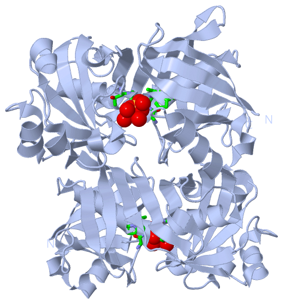
Ligands, Modified Residues, Ions (1, 1)
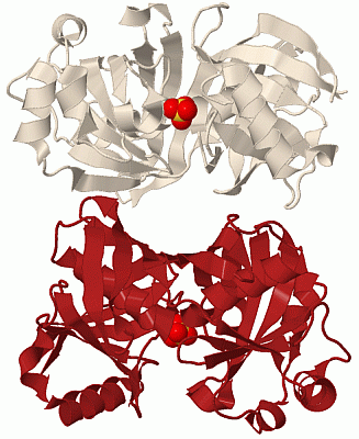
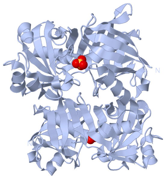
Asymmetric Unit (1, 1)
|
Sites (1, 1)
Asymmetric Unit (1, 1)
|
SS Bonds (0, 0)| (no "SS Bond" information available for 1XUB) |
Cis Peptide Bonds (0, 0)| (no "Cis Peptide Bond" information available for 1XUB) |
SAPs(SNPs)/Variants (0, 0)| (no "SAP(SNP)/Variant" information available for 1XUB) |
PROSITE Motifs (0, 0)| (no "PROSITE Motif" information available for 1XUB) |
Exons (0, 0)| (no "Exon" information available for 1XUB) |
Sequences/Alignments
Asymmetric UnitChain A from PDB Type:PROTEIN Length:278 aligned with PHZF_PSEFL | Q51792 from UniProtKB/Swiss-Prot Length:278 Alignment length:278 10 20 30 40 50 60 70 80 90 100 110 120 130 140 150 160 170 180 190 200 210 220 230 240 250 260 270 PHZF_PSEFL 1 MHNYVIIDAFASVPLEGNPVAVFFDADDLPPAQMQRIAREMNLSESTFVLKPRNGGDALIRIFTPVNELPFAGHPLLGTAIALGAHTDNHRLYLETQMGTIAFELERQNGSVIAASMDQPIPTWTALGRDAELLKALGISDSTFPIEIYHNGPRHVFVGLPSIDALSALHPDHRALSNFHDMAINCFAGAGRRWRSRMFSPAYGVVEDAATGSAAGPLAIHLARHGQIEFGQPVEILQGVEIGRPSLMFAKAEGRAEQLTRVEVSGNGVTFGRGTIVL 278 SCOP domains d1xuba1 A:1-128 Phenazine biosynthesis protein PhzF d1xuba2 A:129-278 Phenazine biosynthesis protein PhzF SCOP domains CATH domains 1xubA01 A:1-119,A:268-278 Diaminopimelate Epimerase; Chain A, domain 1 1xubA02 A:120-267 Diaminopimelate Epimerase; Chain A, domain 1 1xubA01 CATH domains Pfam domains ------PhzC-PhzF-1xubA01 A:7-274 ---- Pfam domains SAPs(SNPs) -------------------------------------------------------------------------------------------------------------------------------------------------------------------------------------------------------------------------------------------------------------------------------------- SAPs(SNPs) PROSITE -------------------------------------------------------------------------------------------------------------------------------------------------------------------------------------------------------------------------------------------------------------------------------------- PROSITE Transcript -------------------------------------------------------------------------------------------------------------------------------------------------------------------------------------------------------------------------------------------------------------------------------------- Transcript 1xub A 1 MHNYVIIDAFASVPLEGNPVAVFFDADDLPPAQMQRIAREMNLSESTFVLKPRNGGDALIRIFTPVNELPFAGAPLLGTAIALGAHTDNHRLYLETQMGTIAFELERQNGSVIAASMDQPIPTWTALGRDAELLKALGISDSTFPIEIYHNGPRHVFVGLPSIDALSALHPDHRALSNFHDMAINCFAGAGRRWRSRMFSPAYGVVEDAATGSAAGPLAIHLARHGQIEFGQPVEILQGVEIGRPSLMFAKAEGRAEQLTRVEVSGNGVTFGRGTIVL 278 10 20 30 40 50 60 70 80 90 100 110 120 130 140 150 160 170 180 190 200 210 220 230 240 250 260 270
|
||||||||||||||||||||
SCOP Domains (1, 2)
Asymmetric Unit
 |
CATH Domains (1, 2)
Asymmetric Unit
|
Pfam Domains (1, 1)| Asymmetric Unit |
Gene Ontology (6, 6)|
Asymmetric Unit(hide GO term definitions) Chain A (PHZF_PSEFL | Q51792)
|
||||||||||||||||||||||||||||||||||||||||||||||||
Interactive Views
|
||||||||||||||||||||||||||||||||||||||||||||||||||||||||||||||||||||||||||||||||||||||||||||||||||||||||||||||||||||||||||||||||||||||||
Still Images
|
||||||||||||||||
Databases
|
||||||||||||||||||||||||||||||||||||||||||||||||||||||||||||||||||||||||||||||||||||||||||||||||||||||||||||||||||||||||||||||||||||||||||||||||||||||||||||||||
Analysis Tools
|
|||||||||||||||||||||||||||||||||||||||||||||||||||||||||||||
Entries Sharing at Least One Protein Chain (UniProt ID)
Related Entries Specified in the PDB File
|
|