|
|
|
|
 Description
|
|
Compounds
|
||||||||||||||||||||||||||||||||||||
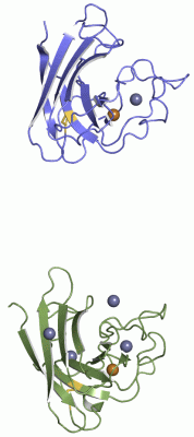
Chains, Units
Summary Information (see also Sequences/Alignments below) |
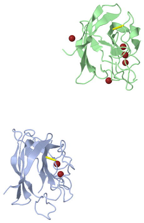
Ligands, Modified Residues, Ions (2, 7)| Asymmetric Unit (2, 7) Biological Unit 1 (0, 0) Biological Unit 2 (0, 0) Biological Unit 3 (0, 0) |
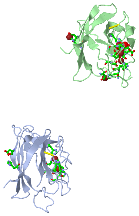
Sites (7, 7)
Asymmetric Unit (7, 7)
|
SS Bonds (2, 2)
Asymmetric Unit
|
||||||||||||
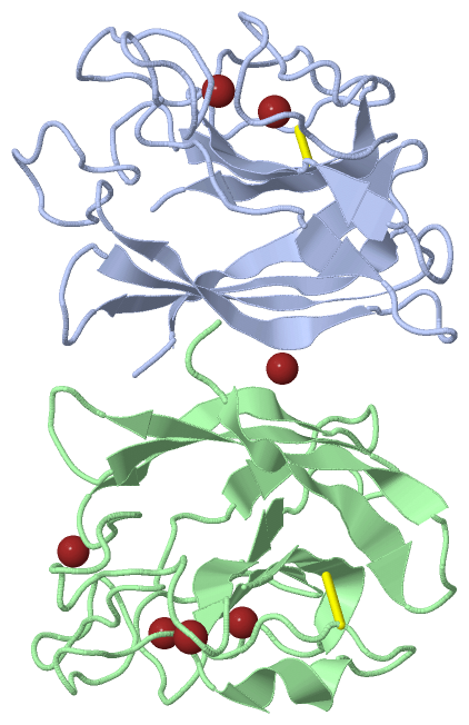
Cis Peptide Bonds (12, 12)
Asymmetric Unit
|
||||||||||||||||||||||||||||||||||||||||||||||||||||
SAPs(SNPs)/Variants (0, 0)| (no "SAP(SNP)/Variant" information available for 1XTM) |
PROSITE Motifs (0, 0)| (no "PROSITE Motif" information available for 1XTM) |
Exons (0, 0)| (no "Exon" information available for 1XTM) |
Sequences/Alignments
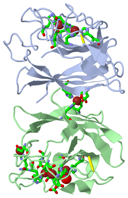
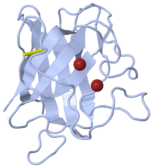
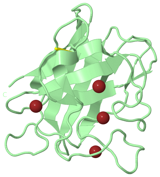
Asymmetric UnitChain A from PDB Type:PROTEIN Length:152 aligned with YOJM_BACSU | O31851 from UniProtKB/Swiss-Prot Length:196 Alignment length:152 49 59 69 79 89 99 109 119 129 139 149 159 169 179 189 YOJM_BACSU 40 AFGHHVQLVNREGKAVGFIEIKESDDEGLDIHISANSLRPGASLGFHIYEKGSCVRPDFESAGGPFNPLNKEHGFNNPMGHHAGDLPNLEVGADGKVDVIMNAPDTSLKKGSKLNILDEDGSAFIIHEQADDYLTNPSGNSGARIVCGALLG 191 SCOP domains d1xtma_ A: automated matches SCOP domains CATH domains -------------------------------------------------------------------------------------------------------------------------------------------------------- CATH domains Pfam domains -------------------------------------------------------------------------------------------------------------------------------------------------------- Pfam domains SAPs(SNPs) -------------------------------------------------------------------------------------------------------------------------------------------------------- SAPs(SNPs) PROSITE -------------------------------------------------------------------------------------------------------------------------------------------------------- PROSITE Transcript -------------------------------------------------------------------------------------------------------------------------------------------------------- Transcript 1xtm A 40 AFGHHVQLVNREGKAVGFIEIKESDDEGLDIHISANSLRPGASLGFHIHEKGSCVRPDFESAGGHFNPLNKEHGFNNPMGHHAGDLPNLEVGADGKVDVIMNAPDTSLKKGSKLNILDEDGSAFIIHEQADDYLTNPSGNSGARIVCGALLG 191 49 59 69 79 89 99 109 119 129 139 149 159 169 179 189 Chain B from PDB Type:PROTEIN Length:153 aligned with YOJM_BACSU | O31851 from UniProtKB/Swiss-Prot Length:196 Alignment length:153 48 58 68 78 88 98 108 118 128 138 148 158 168 178 188 YOJM_BACSU 39 SAFGHHVQLVNREGKAVGFIEIKESDDEGLDIHISANSLRPGASLGFHIYEKGSCVRPDFESAGGPFNPLNKEHGFNNPMGHHAGDLPNLEVGADGKVDVIMNAPDTSLKKGSKLNILDEDGSAFIIHEQADDYLTNPSGNSGARIVCGALLG 191 SCOP domains d1xtmb_ B: automated matches SCOP domains CATH domains --------------------------------------------------------------------------------------------------------------------------------------------------------- CATH domains Pfam domains (1) --Sod_Cu-1xtmB01 B:41-189 -- Pfam domains (1) Pfam domains (2) --Sod_Cu-1xtmB02 B:41-189 -- Pfam domains (2) SAPs(SNPs) --------------------------------------------------------------------------------------------------------------------------------------------------------- SAPs(SNPs) PROSITE --------------------------------------------------------------------------------------------------------------------------------------------------------- PROSITE Transcript --------------------------------------------------------------------------------------------------------------------------------------------------------- Transcript 1xtm B 39 SAFGHHVQLVNREGKAVGFIEIKESDDEGLDIHISANSLRPGASLGFHIHEKGSCVRPDFESAGGHFNPLNKEHGFNNPMGHHAGDLPNLEVGADGKVDVIMNAPDTSLKKGSKLNILDEDGSAFIIHEQADDYLTNPSGNSGARIVCGALLG 191 48 58 68 78 88 98 108 118 128 138 148 158 168 178 188
|
||||||||||||||||||||
SCOP Domains (1, 2)
Asymmetric Unit
 |
CATH Domains (0, 0)| (no "CATH Domain" information available for 1XTM) |
Pfam Domains (1, 2)
Asymmetric Unit
|
Gene Ontology (10, 10)|
Asymmetric Unit(hide GO term definitions) Chain A,B (YOJM_BACSU | O31851)
|
||||||||||||||||||||||||||||||||||||||||||||||||||||||||||||||||||||||||||||||
Interactive Views
|
|||||||||||||||||||||||||||||||||||||||||||||||||||||||||||||||||||||||||||||||||||||||||||||||||||||||||||||||||||||||||||||||||||||||||||||||||||||||||||||||||||||||||||||||||||||||||||||||||||||||||||||||||||||||||||||||||||||||||||||||||||||||||||||||||||||||||||||||||
Still Images
|
||||||||||||||||
Databases
|
||||||||||||||||||||||||||||||||||||||||||||||||||||||||||||||||||||||||||||||||||||||||||||||||||||||||||||||||||||||||||||||||||||||||||||||||||||||||||||||||
Analysis Tools
|
|||||||||||||||||||||||||||||||||||||||||||||||||||||||||||||
Entries Sharing at Least One Protein Chain (UniProt ID)
Related Entries Specified in the PDB File
|
|