Show PDB file:   
         Plain Text   HTML   (compressed file size)
QuickSearch:   
by PDB,NDB,UniProt,PROSITE Code or Search Term(s)  
(-)Asym./Biol. Unit - manually
(-)Asym./Biol. Unit
(-)Asym./Biol. Unit - sites
collapse expand < >
Image Asym./Biol. Unit - manually
Asym./Biol. Unit - manually  (Jmol Viewer)
Image Asym./Biol. Unit
Asym./Biol. Unit  (Jmol Viewer)
Image Asym./Biol. Unit - sites
Asym./Biol. Unit - sites  (Jmol Viewer)

(-) Description

Title :  THE CRYSTAL STRUCTURE OF UDP-GALNAC: POLYPEPTIDE ALPHA-N-ACETYLGALACTOSAMINYLTRANSFERASE-T1
 
Authors :  T. A. Fritz, J. H. Hurley, L. B. Trinh, J. Shiloach, L. A. Tabak
Date :  17 Sep 04  (Deposition) - 26 Oct 04  (Release) - 13 Jul 11  (Revision)
Method :  X-RAY DIFFRACTION
Resolution :  2.50
Chains :  Asym./Biol. Unit :  A
Keywords :  Glycosyltransferase-A (Gt-A), Transferase (Keyword Search: [Gene Ontology, PubMed, Web (Google))
 
Reference :  T. A. Fritz, J. H. Hurley, L. B. Trinh, J. Shiloach, L. A. Tabak
The Beginnings Of Mucin Biosynthesis: The Crystal Structure Of Udp-Galnac:Polypeptide {Alpha}-N-Acetylgalactosaminyltransferase-T1
Proc. Natl. Acad. Sci. Usa V. 101 15307 2004
PubMed-ID: 15486088  |  Reference-DOI: 10.1073/PNAS.0405657101

(-) Compounds

Molecule 1 - POLYPEPTIDE N-ACETYLGALACTOSAMINYLTRANSFERASE 1
    ChainsA
    EC Number2.4.1.41
    EngineeredYES
    Expression SystemPICHIA PASTORIS
    Expression System StrainSMD1168
    Expression System Taxid4922
    FragmentRESIDUES 88-559: INCLUDES CATALYTIC SUBDOMAIN A (RESIDUES 115-225), CATALYTIC SUBDOMAIN B (RESIDUES 285-347), RICIN B-TYPE LECTIN (RESIDUES 429-551)
    GeneGALNT1
    Organism CommonHOUSE MOUSE
    Organism ScientificMUS MUSCULUS
    Organism Taxid10090

 Structural Features

(-) Chains, Units

  1
Asymmetric/Biological Unit A

Summary Information (see also Sequences/Alignments below)

(-) Ligands, Modified Residues, Ions  (4, 6)

Asymmetric/Biological Unit (4, 6)
No.NameCountTypeFull Name
1BMA1Ligand/IonBETA-D-MANNOSE
2CA2Ligand/IonCALCIUM ION
3MN1Ligand/IonMANGANESE (II) ION
4NAG2Ligand/IonN-ACETYL-D-GLUCOSAMINE

(-) Sites  (6, 6)

Asymmetric Unit (6, 6)
No.NameEvidenceResiduesDescription
1AC1SOFTWARETYR A:428 , PHE A:429 , ASN A:552 , NAG A:701BINDING SITE FOR RESIDUE NAG A 700
2AC2SOFTWAREARG A:426 , HIS A:427 , TYR A:428 , NAG A:700 , BMA A:702BINDING SITE FOR RESIDUE NAG A 701
3AC3SOFTWARENAG A:701BINDING SITE FOR RESIDUE BMA A 702
4AC4SOFTWAREASP A:209 , HIS A:211 , HIS A:344BINDING SITE FOR RESIDUE MN A 600
5AC5SOFTWAREASP A:301 , GLN A:304 , GLU A:507 , TYR A:508BINDING SITE FOR RESIDUE CA A 601
6AC6SOFTWARELYS A:512BINDING SITE FOR RESIDUE CA A 602

(-) SS Bonds  (5, 5)

Asymmetric/Biological Unit
No.Residues
1A:106 -A:339
2A:330 -A:408
3A:442 -A:459
4A:482 -A:497
5A:523 -A:540

(-) Cis Peptide Bonds  (0, 0)

(no "Cis Peptide Bond" information available for 1XHB)

 Sequence-Structure Mapping

(-) SAPs(SNPs)/Variants  (0, 0)

(no "SAP(SNP)/Variant" information available for 1XHB)

(-) PROSITE Motifs  (1, 1)

Asymmetric/Biological Unit (1, 1)
 PROSITEUniProtKBPDB
No.IDACDescriptionIDLocationCountLocation
1RICIN_B_LECTINPS50231 Lectin domain of ricin B chain profile.GALT1_MOUSE439-551  1A:439-551

(-) Exons   (0, 0)

(no "Exon" information available for 1XHB)

(-) Sequences/Alignments

Asymmetric/Biological Unit
   Reformat: Number of residues per line =  ('0' or empty: single-line sequence representation)
  Number of residues per labelling interval =   
  UniProt sequence: complete  aligned part    
   Show mapping: SCOP domains CATH domains Pfam domains Secondary structure (by author)
SAPs(SNPs) PROSITE motifs Exons
(details for a mapped element are shown in a popup box when the mouse pointer rests over it)
Chain A from PDB  Type:PROTEIN  Length:447
 aligned with GALT1_MOUSE | O08912 from UniProtKB/Swiss-Prot  Length:559

    Alignment length:459
                                   104       114       124       134       144       154       164       174       184       194       204       214       224       234       244       254       264       274       284       294       304       314       324       334       344       354       364       374       384       394       404       414       424       434       444       454       464       474       484       494       504       514       524       534       544         
          GALT1_MOUSE    95 NRSLPDVRLEGCKTKVYPDNLPTTSVVIVFHNEAWSTLLRTVHSVINRSPRHMIEEIVLVDDASERDFLKRPLESYVKKLKVPVHVIRMEQRSGLIRARLKGAAVSRGQVITFLDAHCECTAGWLEPLLARIKHDRRTVVCPIIDVISDDTFEYMAGSDMTYGGFNWKLNFRWYPVPQREMDRRKGDRTLPVRTPTMAGGLFSIDRDYFQEIGTYDAGMDIWGGENLEISFRIWQCGGTLEIVTCSHVGHVFRKATPYTFPGGTGQIINKNNRRLAEVWMDEFKNFFYIISPGVTKVDYGDISSRLGLRRKLQCKPFSWYLENIYPDSQIPRHYFSLGEIRNVETNQCLDNMARKENEKVGIFNCHGMGGNQVFSYTANKEIRTDDLCLDVSKLNGPVTMLKCHHLKGNQLWEYDPVKLTLQHVNSNQCLDKATEEDSQVPSIRDCTGSRSQQWLLRNV 553
               SCOP domains d1xhba2 A:95-422 Polypeptide N-acetylgalactosaminyltransferase 1, N-terminal domain                                                                                                                                                                                                                                                     d1xhba1 A:423-553 Polypeptide N-acetylgalactosaminyltransferase 1, C-terminal domain                                                SCOP domains
               CATH domains --------------------------------------------------------------------------------------------------------------------------------------------------------------------------------------------------------------------------------------------------------------------------------------------------------------------------------------------------------------------------------------------------------------------------------------------------------------------------- CATH domains
               Pfam domains ------------------------Glycos_transf_2-1xhbA01 A:119-303                                                                                                                                                        ------------------------------------------------------------------------------------------------------------------------------Ricin_B_lectin-1xhbA02 A:430-548                                                                                       ----- Pfam domains
         Sec.struct. author ........hhhhhh.........eeeeeee...hhhhhhhhhhhhhhh.hhh.eeeeeeee....hhhhhhhhhhhhhh....eeeee.....hhhhhhhhhhhhh...eeeeee..eee...hhhhhhhhhhhh..eeeeeeeeee......ee.....eeeee.....eeeee.hhhhhhhh.......ee........eeeehhhhhhh..............hhhhhhhhhh..eeeeeeeeeeeee.------------hhhhhhhhhhhhhhhhhhhhhhhhhh..hhhhh....hhhhhhhhhhh...hhhhhhhhh.........eeeee.eee....eeee.......ee.eeee....hhhhheeee....eee..eeee........eeee....hhhh.eeee....eeee....eeee..........eeee...hhhh.eee... Sec.struct. author
                 SAPs(SNPs) --------------------------------------------------------------------------------------------------------------------------------------------------------------------------------------------------------------------------------------------------------------------------------------------------------------------------------------------------------------------------------------------------------------------------------------------------------------------------- SAPs(SNPs)
                    PROSITE --------------------------------------------------------------------------------------------------------------------------------------------------------------------------------------------------------------------------------------------------------------------------------------------------------------------------------------------------------RICIN_B_LECTIN  PDB: A:439-551 UniProt: 439-551                                                                  -- PROSITE
                 Transcript --------------------------------------------------------------------------------------------------------------------------------------------------------------------------------------------------------------------------------------------------------------------------------------------------------------------------------------------------------------------------------------------------------------------------------------------------------------------------- Transcript
                 1xhb A  95 NRSLPDVRLEGCKTKVYPDNLPTTSVVIVFHNEAWSTLLRTVHSVINRSPRHMIEEIVLVDDASERDFLKRPLESYVKKLKVPVHVIRMEQRSGLIRARLKGAAVSRGQVITFLDAHCECTAGWLEPLLARIKHDRRTVVCPIIDVISDDTFEYMAGSDMTYGGFNWKLNFRWYPVPQREMDRRKGDRTLPVRTPTMAGGLFSIDRDYFQEIGTYDAGMDIWGGENLEISFRIWQCGGTLEIVTCSHVGHVF------------GQIINKNNRRLAEVWMDEFKNFFYIISPGVTKVDYGDISSRLGLRRKLQCKPFSWYLENIYPDSQIPRHYFSLGEIRNVETNQCLDNMARKENEKVGIFNCHGMGGNQVFSYTANKEIRTDDLCLDVSKLNGPVTMLKCHHLKGNQLWEYDPVKLTLQHVNSNQCLDKATEEDSQVPSIRDCTGSRSQQWLLRNV 553
                                   104       114       124       134       144       154       164       174       184       194       204       214       224       234       244       254       264       274       284       294       304       314       324       334       344 |       -    |  364       374       384       394       404       414       424       434       444       454       464       474       484       494       504       514       524       534       544         
                                                                                                                                                                                                                                                                                     346          359                                                                                                                                                                                                  

   Legend:   → Mismatch (orange background)
  - → Gap (green background, '-', border residues have a numbering label)
    → Modified Residue (blue background, lower-case, 'x' indicates undefined single-letter code, labelled with number + name)
  x → Chemical Group (purple background, 'x', labelled with number + name, e.g. ACE or NH2)
  extra numbering lines below/above indicate numbering irregularities and modified residue names etc., number ends below/above '|'

 Classification and Annotation

(-) SCOP Domains  (2, 2)

Asymmetric/Biological Unit

(-) CATH Domains  (0, 0)

(no "CATH Domain" information available for 1XHB)

(-) Pfam Domains  (2, 2)

Asymmetric/Biological Unit
(-)
Clan: GT-A (172)
(-)
Clan: Trefoil (131)

(-) Gene Ontology  (16, 16)

Asymmetric/Biological Unit(hide GO term definitions)
Chain A   (GALT1_MOUSE | O08912)
molecular function
    GO:0030246    carbohydrate binding    Interacting selectively and non-covalently with any carbohydrate, which includes monosaccharides, oligosaccharides and polysaccharides as well as substances derived from monosaccharides by reduction of the carbonyl group (alditols), by oxidation of one or more hydroxy groups to afford the corresponding aldehydes, ketones, or carboxylic acids, or by replacement of one or more hydroxy group(s) by a hydrogen atom. Cyclitols are generally not regarded as carbohydrates.
    GO:0030145    manganese ion binding    Interacting selectively and non-covalently with manganese (Mn) ions.
    GO:0046872    metal ion binding    Interacting selectively and non-covalently with any metal ion.
    GO:0004653    polypeptide N-acetylgalactosaminyltransferase activity    Catalysis of the reaction: UDP-N-acetyl-D-galactosamine + polypeptide = UDP + N-acetyl-D-galactosaminyl-polypeptide. This reaction is the modification of serine or threonine residues in polypeptide chains by the transfer of a N-acetylgalactose from UDP-N-acetylgalactose to the hydroxyl group of the amino acid; it is the first step in O-glycan biosynthesis.
    GO:0016740    transferase activity    Catalysis of the transfer of a group, e.g. a methyl group, glycosyl group, acyl group, phosphorus-containing, or other groups, from one compound (generally regarded as the donor) to another compound (generally regarded as the acceptor). Transferase is the systematic name for any enzyme of EC class 2.
    GO:0016757    transferase activity, transferring glycosyl groups    Catalysis of the transfer of a glycosyl group from one compound (donor) to another (acceptor).
biological process
    GO:0006493    protein O-linked glycosylation    A protein glycosylation process in which a carbohydrate or carbohydrate derivative unit is added to a protein via the hydroxyl group of peptidyl-serine, peptidyl-threonine, peptidyl-hydroxylysine, or peptidyl-hydroxyproline, or via the phenol group of peptidyl-tyrosine, forming an O-glycan.
    GO:0018242    protein O-linked glycosylation via serine    The glycosylation of protein via the O3 atom of peptidyl-serine, forming O3-glycosyl-L-serine; the most common forms are N-acetylgalactosaminyl, mannosyl, galactosyl, and xylosyl serine.
    GO:0018243    protein O-linked glycosylation via threonine    The glycosylation of protein via the O3 atom of peptidyl-threonine, forming O3-glycosyl-L-threonine; the most common forms are N-acetylgalactosaminyl, mannosyl, and galactosyl threonine.
    GO:0006486    protein glycosylation    A protein modification process that results in the addition of a carbohydrate or carbohydrate derivative unit to a protein amino acid, e.g. the addition of glycan chains to proteins.
cellular component
    GO:0005794    Golgi apparatus    A compound membranous cytoplasmic organelle of eukaryotic cells, consisting of flattened, ribosome-free vesicles arranged in a more or less regular stack. The Golgi apparatus differs from the endoplasmic reticulum in often having slightly thicker membranes, appearing in sections as a characteristic shallow semicircle so that the convex side (cis or entry face) abuts the endoplasmic reticulum, secretory vesicles emerging from the concave side (trans or exit face). In vertebrate cells there is usually one such organelle, while in invertebrates and plants, where they are known usually as dictyosomes, there may be several scattered in the cytoplasm. The Golgi apparatus processes proteins produced on the ribosomes of the rough endoplasmic reticulum; such processing includes modification of the core oligosaccharides of glycoproteins, and the sorting and packaging of proteins for transport to a variety of cellular locations. Three different regions of the Golgi are now recognized both in terms of structure and function: cis, in the vicinity of the cis face, trans, in the vicinity of the trans face, and medial, lying between the cis and trans regions.
    GO:0032580    Golgi cisterna membrane    The lipid bilayer surrounding any of the thin, flattened compartments that form the central portion of the Golgi complex.
    GO:0005576    extracellular region    The space external to the outermost structure of a cell. For cells without external protective or external encapsulating structures this refers to space outside of the plasma membrane. This term covers the host cell environment outside an intracellular parasite.
    GO:0016021    integral component of membrane    The component of a membrane consisting of the gene products and protein complexes having at least some part of their peptide sequence embedded in the hydrophobic region of the membrane.
    GO:0016020    membrane    A lipid bilayer along with all the proteins and protein complexes embedded in it an attached to it.
    GO:0048471    perinuclear region of cytoplasm    Cytoplasm situated near, or occurring around, the nucleus.
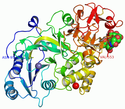
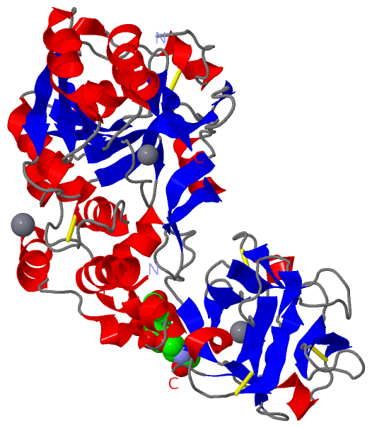
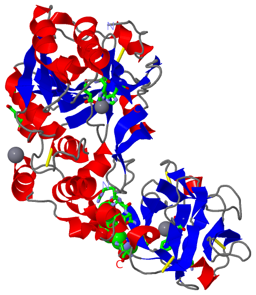
 Visualization

(-) Interactive Views

Asymmetric/Biological Unit
  Complete Structure
    Jena3D(integrated viewing of ligand, site, SAP, PROSITE, SCOP information)
    WebMol | AstexViewer[tm]@PDBe
(Java Applets, require no local installation except for Java; loading may be slow)
    STRAP
(Java WebStart application, automatic local installation, requires Java; full application with system access!)
    RasMol
(require local installation)
    Molscript (VRML)
(requires installation of a VRML viewer; select preferred view via VRML and generate a mono or stereo PDF format file)
 
  Ligands, Modified Residues, Ions
    BMA  [ RasMol | Jena3D ]  +environment [ RasMol | Jena3D ]
    CA  [ RasMol | Jena3D ]  +environment [ RasMol | Jena3D ]
    MN  [ RasMol | Jena3D ]  +environment [ RasMol | Jena3D ]
    NAG  [ RasMol | Jena3D ]  +environment [ RasMol | Jena3D ]
 
  Sites
    AC1  [ RasMol ]  +environment [ RasMol ]
    AC2  [ RasMol ]  +environment [ RasMol ]
    AC3  [ RasMol ]  +environment [ RasMol ]
    AC4  [ RasMol ]  +environment [ RasMol ]
    AC5  [ RasMol ]  +environment [ RasMol ]
    AC6  [ RasMol ]  +environment [ RasMol ]
 
  Cis Peptide Bonds
(no "Cis Peptide Bonds" information available for 1xhb)
 

(-) Still Images

Jmol
  protein: cartoon or spacefill or dots and stick; nucleic acid: cartoon and stick; ligands: spacefill; active site: stick
Molscript
  protein, nucleic acid: cartoon; ligands: spacefill; active site: ball and stick

 Databases and Analysis Tools

(-) Databases

Access by PDB/NDB ID
  1xhb
    Family and Domain InformationProDom | SYSTERS
    General Structural InformationGlycoscienceDB | MMDB | NDB | OCA | PDB | PDBe | PDBj | PDBsum | PDBWiki | PQS | PROTEOPEDIA
    Orientation in MembranesOPM
    Protein SurfaceSURFACE
    Secondary StructureDSSP (structure derived) | HSSP (homology derived)
    Structural GenomicsGeneCensus
    Structural NeighboursCE | VAST
    Structure ClassificationCATH | Dali | SCOP
    Validation and Original DataBMRB Data View | BMRB Restraints Grid | EDS | PROCHECK | RECOORD | WHAT_CHECK
 
Access by UniProt ID/Accession number
  GALT1_MOUSE | O08912
    Comparative Protein Structure ModelsModBase
    Genomic InformationEnsembl
    Protein-protein InteractionDIP
    Sequence, Family and Domain InformationInterPro | Pfam | SMART | UniProtKB/SwissProt
 
Access by Enzyme Classificator   (EC Number)
  2.4.1.41
    General Enzyme InformationBRENDA | EC-PDB | Enzyme | IntEnz
    PathwayKEGG | MetaCyc
 
Access by Disease Identifier   (MIM ID)
  (no 'MIM ID' available)
    Disease InformationOMIM
 
Access by GenAge ID
  (no 'GenAge ID' available)
    Age Related InformationGenAge

(-) Analysis Tools

Access by PDB/NDB ID
    Domain InformationXDom
    Interatomic Contacts of Structural UnitsCSU
    Ligand-protein ContactsLPC
    Protein CavitiescastP
    Sequence and Secondary StructurePDBCartoon
    Structure AlignmentSTRAP(Java WebStart application, automatic local installation, requires Java; full application with system access!)
    Structure and Sequence BrowserSTING
 
Access by UniProt ID/Accession number
  GALT1_MOUSE | O08912
    Protein Disorder PredictionDisEMBL | FoldIndex | GLOBPLOT (for more information see DisProt)

 Related Entries

(-) Entries Sharing at Least One Protein Chain (UniProt ID)

(no "Entries Sharing at Least One Protein Chain" available for 1XHB)

(-) Related Entries Specified in the PDB File

(no "Related Entries Specified in the PDB File" available for 1XHB)