Show PDB file:   
         Plain Text   HTML   (compressed file size)
QuickSearch:   
by PDB,NDB,UniProt,PROSITE Code or Search Term(s)  
(-)NMR Structure - model 1
(-)NMR Structure - model 1, sites
(-)NMR Structure - all models
collapse expand < >
Image NMR Structure - model 1
NMR Structure - model 1  (Jmol Viewer)
Image NMR Structure - model 1, sites
NMR Structure - model 1, sites  (Jmol Viewer)
Image NMR Structure - all models
NMR Structure - all models  (Jmol Viewer)

(-) Description

Title :  SOLUTION STRUCTURE OF THE FYVE DOMAIN FROM HUMAN FYVE DOMAIN CONTAINING 27 ISOFORM B PROTEIN
 
Authors :  T. Tomizawa, T. Kigawa, S. Koshiba, M. Inoue, S. Yokoyama, Riken Structural Genomics/Proteomics Initiative (Rsgi)
Date :  15 May 05  (Deposition) - 15 Nov 05  (Release) - 24 Feb 09  (Revision)
Method :  SOLUTION NMR
Resolution :  NOT APPLICABLE
Chains :  NMR Structure  :  A  (20x)
Keywords :  Fyve Domain, Phosphoinositide Binding, Zinc Binding, Structural Genomics, Nppsfa, National Project On Protein Structural And Functional Analyses, Riken Structural Genomics/Proteomics Initiative, Rsgi, Lipid Binding Protein (Keyword Search: [Gene Ontology, PubMed, Web (Google))
 
Reference :  T. Tomizawa, T. Kigawa, S. Koshiba, M. Inoue, S. Yokoyama
Solution Structure Of The Fyve Domain From Human Fyve Domain Containing 27 Isoform B Protein
To Be Published
PubMed: search
(for further references see the PDB file header)

(-) Compounds

Molecule 1 - ZINC FINGER, FYVE DOMAIN CONTAINING 27 ISOFORM B
    ChainsA
    EngineeredYES
    Expression System PlasmidP041206-02
    Expression System Vector TypePLASMID
    FragmentFYVE DOMAIN
    GeneZFYVE27
    Organism CommonHUMAN
    Organism ScientificHOMO SAPIENS
    Organism Taxid9606
    Other DetailsCELL-FREE PROTEIN SYNTHESIS

 Structural Features

(-) Chains, Units

  
NMR Structure (20x)

Summary Information (see also Sequences/Alignments below)

(-) Ligands, Modified Residues, Ions  (1, 2)

NMR Structure (1, 2)
No.NameCountTypeFull Name
1ZN2Ligand/IonZINC ION

(-) Sites  (2, 2)

NMR Structure (2, 2)
No.NameEvidenceResiduesDescription
1AC1SOFTWARECYS A:17 , CYS A:20 , CYS A:41 , CYS A:44BINDING SITE FOR RESIDUE ZN A 201
2AC2SOFTWARECYS A:33 , CYS A:36 , PHE A:40 , CYS A:69 , CYS A:72BINDING SITE FOR RESIDUE ZN A 401

(-) SS Bonds  (0, 0)

(no "SS Bond" information available for 1X4U)

(-) Cis Peptide Bonds  (0, 0)

(no "Cis Peptide Bond" information available for 1X4U)

 Sequence-Structure Mapping

(-) SAPs(SNPs)/Variants  (0, 0)

(no "SAP(SNP)/Variant" information available for 1X4U)

(-) PROSITE Motifs  (1, 1)

NMR Structure (1, 1)
 PROSITEUniProtKBPDB
No.IDACDescriptionIDLocationCountLocation
1ZF_FYVEPS50178 Zinc finger FYVE/FYVE-related type profile.ZFY27_HUMAN344-410  1A:11-77

(-) Exons   (4, 4)

NMR Structure (4, 4)
 ENSEMBLUniProtKBPDB
No.Transcript IDExonExon IDGenome LocationLengthIDLocationLengthCountLocationLength
1.1bENST000003936771bENSE00001955102chr10:99496880-99497082203ZFY27_HUMAN-00--
1.2bENST000003936772bENSE00002191420chr10:99498234-99498431198ZFY27_HUMAN1-66660--
1.3ENST000003936773ENSE00001713838chr10:99502851-9950292171ZFY27_HUMAN66-90250--
1.4ENST000003936774ENSE00001019956chr10:99504486-99504672187ZFY27_HUMAN90-152630--
1.5ENST000003936775ENSE00001019964chr10:99508026-9950812196ZFY27_HUMAN152-184330--
1.6ENST000003936776ENSE00001019965chr10:99509231-99509343113ZFY27_HUMAN184-222390--
1.7bENST000003936777bENSE00001019961chr10:99510088-99510227140ZFY27_HUMAN222-268470--
1.8cENST000003936778cENSE00001019974chr10:99511148-9951121972ZFY27_HUMAN269-292240--
1.9ENST000003936779ENSE00001236963chr10:99512614-9951263421ZFY27_HUMAN293-29970--
1.10ENST0000039367710ENSE00001097766chr10:99512795-99512939145ZFY27_HUMAN300-348491A:1-15 (gaps)45
1.12cENST0000039367712cENSE00001236979chr10:99517022-9951706847ZFY27_HUMAN348-363161A:15-3016
1.13ENST0000039367713ENSE00001236974chr10:99517399-9951748082ZFY27_HUMAN364-391281A:31-5828
1.15bENST0000039367715bENSE00001389121chr10:99518993-995206521660ZFY27_HUMAN391-411211A:58-7821

(-) Sequences/Alignments

NMR Structure
   Reformat: Number of residues per line =  ('0' or empty: single-line sequence representation)
  Number of residues per labelling interval =   
  UniProt sequence: complete  aligned part    
   Show mapping: SCOP domains CATH domains Pfam domains Secondary structure (by author)
SAPs(SNPs) PROSITE motifs Exons
(details for a mapped element are shown in a popup box when the mouse pointer rests over it)
Chain A from PDB  Type:PROTEIN  Length:84
 aligned with ZFY27_HUMAN | Q5T4F4 from UniProtKB/Swiss-Prot  Length:411

    Alignment length:114
                                                                                                                                     411      
                                   313       323       333       343       353       363       373       383       393       403       | -    
          ZFY27_HUMAN   304 GAPCPAEDELALQDNGFLSKNEVLRSKVSRLTERLRKRYPTNNFGNCTGCSATFSVLKKRRSCSNCGNSFCSRCCSFKVPKSSMGATAPEAQRETVFVCASCNQTLSK------   -
               SCOP domains ------------------------------------------------------------------------------------------------------------------ SCOP domains
               CATH domains ------------------------------------------------------------------------------------------------------------------ CATH domains
               Pfam domains -------------------------------------FYVE-1x4uA01 A:8-78                                                    ------ Pfam domains
         Sec.struct. author ..-----------------------....-------........................ee......ee......eee................eeehhhhhhhhhh...... Sec.struct. author
                 SAPs(SNPs) ------------------------------------------------------------------------------------------------------------------ SAPs(SNPs)
                    PROSITE ----------------------------------------ZF_FYVE  PDB: A:11-77 UniProt: 344-410                             ------- PROSITE
           Transcript 1 (1) Exon 1.10  PDB: A:1-15 (gaps) [INCOMPLETE]   ---------------Exon 1.13  PDB: A:31-58     -------------------------- Transcript 1 (1)
           Transcript 1 (2) --------------------------------------------Exon 1.12c      ---------------------------Exon 1.15b           ------ Transcript 1 (2)
                 1x4u A   1 GS-----------------------SGSS-------GRYPTNNFGNCTGCSATFSVLKKRRSCSNCGNSFCSRCCSFKVPKSSMGATAPEAQRETVFVCASCNQTLSKSGPSSG  84
                             |       -         -     |  |-      | 10        20        30        40        50        60        70        80    
                             2                       3  6       7                                                                             

   Legend:   → Mismatch (orange background)
  - → Gap (green background, '-', border residues have a numbering label)
    → Modified Residue (blue background, lower-case, 'x' indicates undefined single-letter code, labelled with number + name)
  x → Chemical Group (purple background, 'x', labelled with number + name, e.g. ACE or NH2)
  extra numbering lines below/above indicate numbering irregularities and modified residue names etc., number ends below/above '|'

 Classification and Annotation

(-) SCOP Domains  (0, 0)

(no "SCOP Domain" information available for 1X4U)

(-) CATH Domains  (0, 0)

(no "CATH Domain" information available for 1X4U)

(-) Pfam Domains  (1, 1)

NMR Structure

(-) Gene Ontology  (18, 18)

NMR Structure(hide GO term definitions)
Chain A   (ZFY27_HUMAN | Q5T4F4)
molecular function
    GO:0046872    metal ion binding    Interacting selectively and non-covalently with any metal ion.
    GO:0005515    protein binding    Interacting selectively and non-covalently with any protein or protein complex (a complex of two or more proteins that may include other nonprotein molecules).
biological process
    GO:0031175    neuron projection development    The process whose specific outcome is the progression of a neuron projection over time, from its formation to the mature structure. A neuron projection is any process extending from a neural cell, such as axons or dendrites (collectively called neurites).
    GO:0048011    neurotrophin TRK receptor signaling pathway    A series of molecular signals initiated by the binding of a neurotrophin to a receptor on the surface of the target cell where the receptor possesses tyrosine kinase activity, and ending with regulation of a downstream cellular process, e.g. transcription.
    GO:0072659    protein localization to plasma membrane    A process in which a protein is transported to, or maintained in, a specific location in the plasma membrane.
cellular component
    GO:0030424    axon    The long process of a neuron that conducts nerve impulses, usually away from the cell body to the terminals and varicosities, which are sites of storage and release of neurotransmitter.
    GO:0042995    cell projection    A prolongation or process extending from a cell, e.g. a flagellum or axon.
    GO:0005737    cytoplasm    All of the contents of a cell excluding the plasma membrane and nucleus, but including other subcellular structures.
    GO:0030425    dendrite    A neuron projection that has a short, tapering, often branched, morphology, receives and integrates signals from other neurons or from sensory stimuli, and conducts a nerve impulse towards the axon or the cell body. In most neurons, the impulse is conveyed from dendrites to axon via the cell body, but in some types of unipolar neuron, the impulse does not travel via the cell body.
    GO:0005783    endoplasmic reticulum    The irregular network of unit membranes, visible only by electron microscopy, that occurs in the cytoplasm of many eukaryotic cells. The membranes form a complex meshwork of tubular channels, which are often expanded into slitlike cavities called cisternae. The ER takes two forms, rough (or granular), with ribosomes adhering to the outer surface, and smooth (with no ribosomes attached).
    GO:0005789    endoplasmic reticulum membrane    The lipid bilayer surrounding the endoplasmic reticulum.
    GO:0071782    endoplasmic reticulum tubular network    A subcompartment of the endoplasmic reticulum consisting of tubules having membranes with high curvature in cross-section.
    GO:0005768    endosome    A vacuole to which materials ingested by endocytosis are delivered.
    GO:0032584    growth cone membrane    The portion of the plasma membrane surrounding a growth cone.
    GO:0016021    integral component of membrane    The component of a membrane consisting of the gene products and protein complexes having at least some part of their peptide sequence embedded in the hydrophobic region of the membrane.
    GO:0016020    membrane    A lipid bilayer along with all the proteins and protein complexes embedded in it an attached to it.
    GO:0005886    plasma membrane    The membrane surrounding a cell that separates the cell from its external environment. It consists of a phospholipid bilayer and associated proteins.
    GO:0055038    recycling endosome membrane    The lipid bilayer surrounding a recycling endosome.
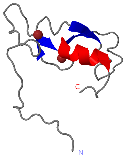
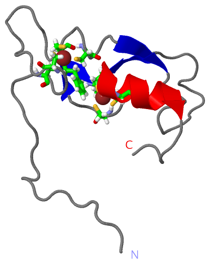
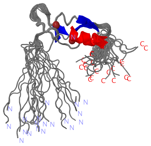
 Visualization

(-) Interactive Views

NMR Structure
  Complete Structure
    Jena3D(integrated viewing of ligand, site, SAP, PROSITE, SCOP information)
    WebMol | AstexViewer[tm]@PDBe
(Java Applets, require no local installation except for Java; loading may be slow)
    STRAP
(Java WebStart application, automatic local installation, requires Java; full application with system access!)
    RasMol
(require local installation)
    Molscript (VRML)
(requires installation of a VRML viewer; select preferred view via VRML and generate a mono or stereo PDF format file)
 
  Ligands, Modified Residues, Ions
    ZN  [ RasMol | Jena3D ]  +environment [ RasMol | Jena3D ]
 
  Sites
    AC1  [ RasMol ]  +environment [ RasMol ]
    AC2  [ RasMol ]  +environment [ RasMol ]
 
  Cis Peptide Bonds
(no "Cis Peptide Bonds" information available for 1x4u)
 

(-) Still Images

Jmol
  protein: cartoon or spacefill or dots and stick; nucleic acid: cartoon and stick; ligands: spacefill; active site: stick
Molscript
  protein, nucleic acid: cartoon; ligands: spacefill; active site: ball and stick

 Databases and Analysis Tools

(-) Databases

Access by PDB/NDB ID
  1x4u
    Family and Domain InformationProDom | SYSTERS
    General Structural InformationGlycoscienceDB | MMDB | NDB | OCA | PDB | PDBe | PDBj | PDBsum | PDBWiki | PQS | PROTEOPEDIA
    Orientation in MembranesOPM
    Protein SurfaceSURFACE
    Secondary StructureDSSP (structure derived) | HSSP (homology derived)
    Structural GenomicsGeneCensus
    Structural NeighboursCE | VAST
    Structure ClassificationCATH | Dali | SCOP
    Validation and Original DataBMRB Data View | BMRB Restraints Grid | EDS | PROCHECK | RECOORD | WHAT_CHECK
 
Access by UniProt ID/Accession number
  ZFY27_HUMAN | Q5T4F4
    Comparative Protein Structure ModelsModBase
    Genomic InformationEnsembl
    Protein-protein InteractionDIP
    Sequence, Family and Domain InformationInterPro | Pfam | SMART | UniProtKB/SwissProt
 
Access by Enzyme Classificator   (EC Number)
  (no 'Enzyme Classificator' available)
    General Enzyme InformationBRENDA | EC-PDB | Enzyme | IntEnz
    PathwayKEGG | MetaCyc
 
Access by Disease Identifier   (MIM ID)
  (no 'MIM ID' available)
    Disease InformationOMIM
 
Access by GenAge ID
  (no 'GenAge ID' available)
    Age Related InformationGenAge

(-) Analysis Tools

Access by PDB/NDB ID
    Domain InformationXDom
    Interatomic Contacts of Structural UnitsCSU
    Ligand-protein ContactsLPC
    Protein CavitiescastP
    Sequence and Secondary StructurePDBCartoon
    Structure AlignmentSTRAP(Java WebStart application, automatic local installation, requires Java; full application with system access!)
    Structure and Sequence BrowserSTING
 
Access by UniProt ID/Accession number
  ZFY27_HUMAN | Q5T4F4
    Protein Disorder PredictionDisEMBL | FoldIndex | GLOBPLOT (for more information see DisProt)

 Related Entries

(-) Entries Sharing at Least One Protein Chain (UniProt ID)

(no "Entries Sharing at Least One Protein Chain" available for 1X4U)

(-) Related Entries Specified in the PDB File

(no "Related Entries Specified in the PDB File" available for 1X4U)