|
|
|
|
 Description
|
|
Compounds
|
||||||||||||||||||||||||||||||||||||||||||||||||||||
Chains, Units
Summary Information (see also Sequences/Alignments below) |
Ligands, Modified Residues, Ions (0, 0)| (no "Ligand,Modified Residues,Ions" information available for 1WUZ) |
Sites (0, 0)| (no "Site" information available for 1WUZ) |
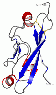
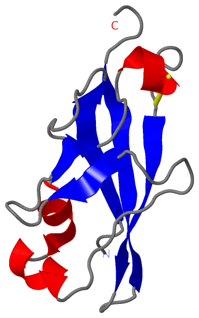
SS Bonds (1, 1)
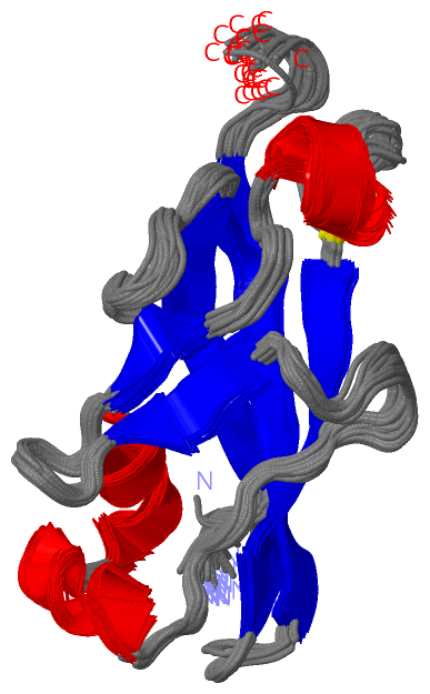
NMR Structure
|
||||||||
Cis Peptide Bonds (1, 30)
NMR Structure
|
||||||||||
SAPs(SNPs)/Variants (0, 0)| (no "SAP(SNP)/Variant" information available for 1WUZ) |
PROSITE Motifs (2, 2)
NMR Structure (2, 2)
|
||||||||||||||||||||||||||||||||
Exons (0, 0)| (no "Exon" information available for 1WUZ) |
Sequences/Alignments
NMR StructureChain A from PDB Type:PROTEIN Length:103 aligned with PCDA4_MOUSE | O88689 from UniProtKB/Swiss-Prot Length:947 Alignment length:103 36 46 56 66 76 86 96 106 116 126 PCDA4_MOUSE 27 GNSQIHYSIPEEAKHGTFVGRIAQDLGLELTELVPRLFRVASKDRGDLLEVNLQNGILFVNSRIDREELCGRSAECSIHLEVIVDRPLQVFHVEVEVRDINDN 129 SCOP domains ------------------------------------------------------------------------------------------------------- SCOP domains CATH domains ------------------------------------------------------------------------------------------------------- CATH domains Pfam domains --Cadherin_2-1wuzA01 A:3-86 ----------------- Pfam domains SAPs(SNPs) ------------------------------------------------------------------------------------------------------- SAPs(SNPs) PROSITE (1) -CADHERIN_2 PDB: A:2-103 UniProt: 28-133 PROSITE (1) PROSITE (2) ----------------------------------------------------------------------------------------------CADHERIN_ PROSITE (2) Transcript ------------------------------------------------------------------------------------------------------- Transcript 1wuz A 1 GNSQIHYSIPEEAKHGTFVGRIAQDLGLELTELVPRLFRVASKDRGDLLEVNLQNGILFVNSRIDREELCGRSAECSIHLEVIVDRPLQVFHVEVEVRDINDN 103 10 20 30 40 50 60 70 80 90 100
|
||||||||||||||||||||
SCOP Domains (0, 0)| (no "SCOP Domain" information available for 1WUZ) |
CATH Domains (0, 0)| (no "CATH Domain" information available for 1WUZ) |
Pfam Domains (1, 1)
NMR Structure

|
Gene Ontology (9, 9)|
NMR Structure(hide GO term definitions) Chain A (PCDA4_MOUSE | O88689)
|
||||||||||||||||||||||||||||||||||||||||||||||||||||||||||||||||||||||||
Interactive Views
|
|||||||||||||||||||||||||||||||||||||||||||||||||||||||||||||||||||||||||||||||||||||||||||||||||||||||||||||||||||||
Still Images
|
||||||||||||||||
Databases
|
||||||||||||||||||||||||||||||||||||||||||||||||||||||||||||||||||||||||||||||||||||||||||||||||||||||||||||||||||||||||||||||||||||||||||||||||||||||||||||||||
Analysis Tools
|
|||||||||||||||||||||||||||||||||||||||||||||||||||||||||||||
Entries Sharing at Least One Protein Chain (UniProt ID)
Related Entries Specified in the PDB File
|
|