|
|
|
|
 Description
|
|
Compounds
|
||||||||||||||||||||||||
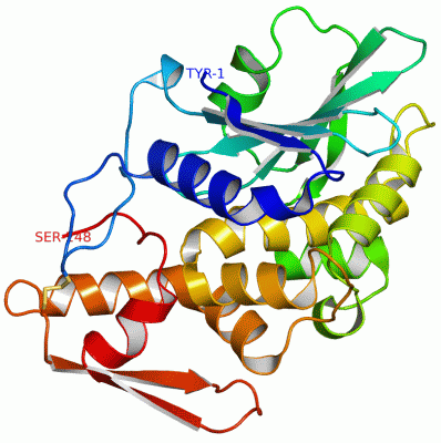
Chains, Units
Summary Information (see also Sequences/Alignments below) |
Ligands, Modified Residues, Ions (0, 0)| (no "Ligand,Modified Residues,Ions" information available for 3CTK) |
Sites (0, 0)| (no "Site" information available for 3CTK) |
SS Bonds (1, 1)
Asymmetric/Biological Unit
|
||||||||
Cis Peptide Bonds (2, 2)
Asymmetric/Biological Unit
|
||||||||||||
SAPs(SNPs)/Variants (0, 0)| (no "SAP(SNP)/Variant" information available for 3CTK) |
PROSITE Motifs (0, 0)| (no "PROSITE Motif" information available for 3CTK) |
Exons (0, 0)| (no "Exon" information available for 3CTK) |
Sequences/Alignments
Asymmetric/Biological UnitChain A from PDB Type:PROTEIN Length:248 aligned with Q8W4U4_9CARY | Q8W4U4 from UniProtKB/TrEMBL Length:305 Alignment length:248 36 46 56 66 76 86 96 106 116 126 136 146 156 166 176 186 196 206 216 226 236 246 256 266 Q8W4U4_9CARY 27 YNTVSFNLGEAYEYPTFIQDLRNELAKGTPVCQLPVTLQTIADDKRFVLVDITTTSKKTVKVAIDVTDVYVVGYQDKWDGKDRAVFLDKVPTVATSKLFPGVTNRVTLTFDGSYQKLVNAAKVDRKDLELGVYKLEFSIEAIHGKTINGQEIAKFFLIVIQMVSEAARFKYIETEVVDRGLYGSFKPNFKVLNLENNWGDISDAIHKSSPQCTTINPALQLISPSNDPWVVNKVSQISPDMGILKFKS 274 SCOP domains --d3ctka1 A:3-248 Bouganin SCOP domains CATH domains 3ctkA01 A:1-167 Ricin (A subunit), domain 1 3ctkA02 A:168-248 Ricin (A Subunit), domain 2 CATH domains Pfam domains -------------------------------------------------------------------------------------------------------------------------------------------------------------------------------------------------------------------------------------------------------- Pfam domains SAPs(SNPs) -------------------------------------------------------------------------------------------------------------------------------------------------------------------------------------------------------------------------------------------------------- SAPs(SNPs) PROSITE -------------------------------------------------------------------------------------------------------------------------------------------------------------------------------------------------------------------------------------------------------- PROSITE Transcript -------------------------------------------------------------------------------------------------------------------------------------------------------------------------------------------------------------------------------------------------------- Transcript 3ctk A 1 YNTVSFNLGEAYEYPTFIQDLRNELAKGTPVCQLPVTLQTIADDKRFVLVDITTTSKKTVKVAIDVTDVYVVGYQDKWDGKDRAVFLDKVPTVATSKLFPGVTNRVTLTFDGSYQKLVNAAKVDRKDLELGVYKLEFSIEAIHGKTINGQEIAKFFLIVIQMVSEAARFKYIETEVVDRGLYGSFKPNFKVLNLENNWGDISDAIHKSSPQCTTINPALQLISPSNDPWVVNKVSQISPDMGILKFKS 248 10 20 30 40 50 60 70 80 90 100 110 120 130 140 150 160 170 180 190 200 210 220 230 240
|
||||||||||||||||||||
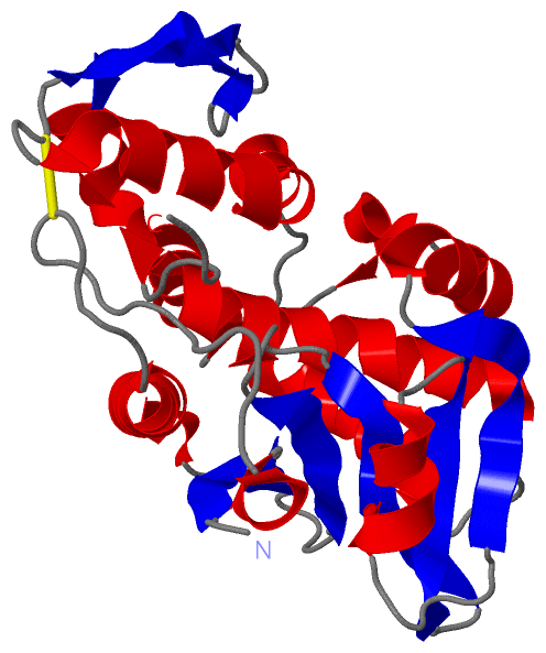
SCOP Domains (1, 1)
Asymmetric/Biological Unit
 |
CATH Domains (2, 2)
Asymmetric/Biological Unit
|
Pfam Domains (0, 0)| (no "Pfam Domain" information available for 3CTK) |
Gene Ontology (4, 4)|
Asymmetric/Biological Unit(hide GO term definitions) Chain A (Q8W4U4_9CARY | Q8W4U4)
|
||||||||||||||||||||||||||||||||||||
Interactive Views
|
||||||||||||||||||||||||||||||||||||||||||||||||||||||||||||||||||||||||||||||||||||||||||||||||||||||||||||||||||||||||||||
Still Images
|
||||||||||||||||
Databases
|
||||||||||||||||||||||||||||||||||||||||||||||||||||||||||||||||||||||||||||||||||||||||||||||||||||||||||||||||||||||||||||||||||||||||||||||||||||||||||||||||
Analysis Tools
|
|||||||||||||||||||||||||||||||||||||||||||||||||||||||||||||
Entries Sharing at Least One Protein Chain (UniProt ID)
Related Entries Specified in the PDB File
|
|