|
|
|
|
 Description
|
|
Compounds
|
||||||||||||||||||||||||||||||||||||||||||||
Chains, Units
Summary Information (see also Sequences/Alignments below) |
Ligands, Modified Residues, Ions (0, 0)| (no "Ligand,Modified Residues,Ions" information available for 1WK1) |
Sites (0, 0)| (no "Site" information available for 1WK1) |
SS Bonds (2, 2)
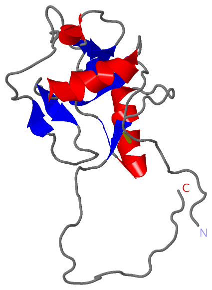
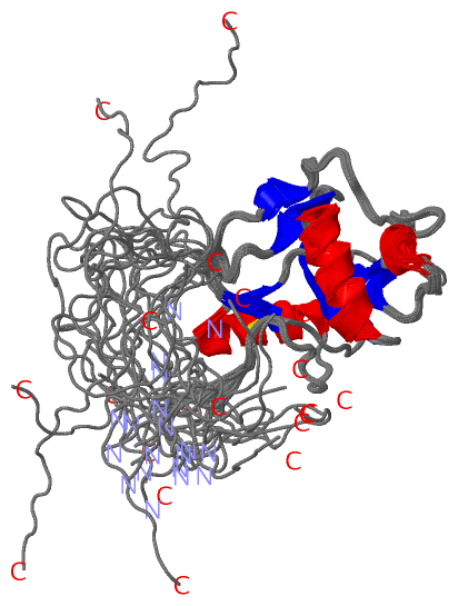
NMR Structure
|
||||||||||||
Cis Peptide Bonds (1, 20)
NMR Structure
|
||||||||||
SAPs(SNPs)/Variants (0, 0)| (no "SAP(SNP)/Variant" information available for 1WK1) |
PROSITE Motifs (0, 0)| (no "PROSITE Motif" information available for 1WK1) |
Exons (0, 0)| (no "Exon" information available for 1WK1) |
Sequences/Alignments
NMR StructureChain A from PDB Type:PROTEIN Length:150 aligned with Q19853_CAEEL | Q19853 from UniProtKB/TrEMBL Length:2229 Alignment length:182 1195 1205 1215 1225 1235 1245 1255 1265 1275 1285 1295 1305 1315 1325 1335 1345 1355 1365 Q19853_CAEEL 1186 ESFGQYCVKFLTVNDDILSMPQARNFCASAGGYLADDLGDDKNNFYSSIAANTQFWIGLFKNSDGQFYWDRGQGINPDLLNQPITYWANGEPSNDPTRQCVYFDGRSGDKSKVWTTDTCATPRPFICQKHRYDSDHKPNTIGDADLPAGDWYVKIKTNPTNSNPPYCSLSVRVQSSLQIVSG 1367 SCOP domains d1wk1a_ A: Hypothetical protein F28B4.3 SCOP domains CATH domains -------------------------------------------------------------------------------------------------------------------------------------------------------------------------------------- CATH domains Pfam domains -----------------Lectin_C-1wk1A01 A:18-129 ----------------------------------------------------- Pfam domains SAPs(SNPs) -------------------------------------------------------------------------------------------------------------------------------------------------------------------------------------- SAPs(SNPs) PROSITE -------------------------------------------------------------------------------------------------------------------------------------------------------------------------------------- PROSITE Transcript -------------------------------------------------------------------------------------------------------------------------------------------------------------------------------------- Transcript 1wk1 A 1 GSSGSSGVKFLTVNDDILSMPQARNFCASAGGYLADDLGDDKNNFYSSIAANTQFWIGLFKNSDGQFYWDRGQGINPDLLNQPITYWANGEPSNDPTRQCVYFDGRSGDKSKVWTTDTCATPRPFICQKHRYDSDHKPNTIGDASGPS--------------------------------SG 150 10 20 30 40 50 60 70 80 90 100 110 120 130 140 | - - - -| 148 149
|
||||||||||||||||||||
SCOP Domains (1, 1)
NMR Structure
 |
CATH Domains (0, 0)| (no "CATH Domain" information available for 1WK1) |
Pfam Domains (1, 1)
NMR Structure
|
Gene Ontology (0, 0)|
NMR Structure(hide GO term definitions)
(no "Gene Ontology" information available for 1WK1)
|
Interactive Views
|
|||||||||||||||||||||||||||||||||||||||||||||||||||||||||||||||||||||||||||||||||||||||||||||||||||||||||||||||||||||
Still Images
|
||||||||||||||||
Databases
|
||||||||||||||||||||||||||||||||||||||||||||||||||||||||||||||||||||||||||||||||||||||||||||||||||||||||||||||||||||||||||||||||||||||||||||||||||||||||||||||||
Analysis Tools
|
|||||||||||||||||||||||||||||||||||||||||||||||||||||||||||||
Entries Sharing at Least One Protein Chain (UniProt ID)
Related Entries Specified in the PDB File
|
|