Show PDB file:   
         Plain Text   HTML   (compressed file size)
QuickSearch:   
by PDB,NDB,UniProt,PROSITE Code or Search Term(s)  
(-)Asym./Biol. Unit
(-)Asym./Biol. Unit - sites
collapse expand < >
Image Asym./Biol. Unit
Asym./Biol. Unit  (Jmol Viewer)
Image Asym./Biol. Unit - sites
Asym./Biol. Unit - sites  (Jmol Viewer)

(-) Description

Title :  SCALLOP MYOSIN REGULATORY DOMAIN
 
Authors :  A. Houdusse, C. Cohen
Date :  19 Jan 96  (Deposition) - 11 Jul 96  (Release) - 24 Feb 09  (Revision)
Method :  X-RAY DIFFRACTION
Resolution :  2.00
Chains :  Asym./Biol. Unit :  A,B,C
Keywords :  Myosin, Calcium Binding Protein, Muscle Protein (Keyword Search: [Gene Ontology, PubMed, Web (Google))
 
Reference :  A. Houdusse, C. Cohen
Structure Of The Regulatory Domain Of Scallop Myosin At 2 A Resolution: Implications For Regulation.
Structure V. 4 21 1996
PubMed-ID: 8805510  |  Reference-DOI: 10.1016/S0969-2126(96)00154-2
(for further references see the PDB file header)

(-) Compounds

Molecule 1 - SCALLOP MYOSIN
    ChainsA
    FragmentPROTEOLYTIC FRAGMENT, REGULATORY DOMAIN
    OrganSKELETAL
    Organism ScientificARGOPECTEN IRRADIANS
    Organism Taxid31199
    Other DetailsPH 7.0
    TissueSKELETAL MUSCLE
 
Molecule 2 - SCALLOP MYOSIN
    ChainsB
    FragmentPROTEOLYTIC FRAGMENT, REGULATORY DOMAIN
    OrganSKELETAL
    Organism ScientificARGOPECTEN IRRADIANS
    Organism Taxid31199
    Other DetailsPH 7.0
    TissueSKELETAL MUSCLE
 
Molecule 3 - SCALLOP MYOSIN
    ChainsC
    FragmentPROTEOLYTIC FRAGMENT, REGULATORY DOMAIN
    OrganSKELETAL
    Organism ScientificARGOPECTEN IRRADIANS
    Organism Taxid31199
    Other DetailsPH 7.0
    TissueSKELETAL MUSCLE

 Structural Features

(-) Chains, Units

  123
Asymmetric/Biological Unit ABC

Summary Information (see also Sequences/Alignments below)

(-) Ligands, Modified Residues, Ions  (2, 2)

Asymmetric/Biological Unit (2, 2)
No.NameCountTypeFull Name
1CA1Ligand/IonCALCIUM ION
2MG1Ligand/IonMAGNESIUM ION

(-) Sites  (2, 2)

Asymmetric Unit (2, 2)
No.NameEvidenceResiduesDescription
1AC1SOFTWAREASP C:19 , ASP C:22 , GLY C:23 , ASP C:25 , ALA C:27 , HOH C:512BINDING SITE FOR RESIDUE CA C 501
2AC2SOFTWAREASP B:28 , ASP B:30 , ASP B:32 , PHE B:34 , ASP B:39 , HOH B:527BINDING SITE FOR RESIDUE MG B 502

(-) SS Bonds  (0, 0)

(no "SS Bond" information available for 1WDC)

(-) Cis Peptide Bonds  (0, 0)

(no "Cis Peptide Bond" information available for 1WDC)

 Sequence-Structure Mapping

(-) SAPs(SNPs)/Variants  (0, 0)

(no "SAP(SNP)/Variant" information available for 1WDC)

(-) PROSITE Motifs  (3, 6)

Asymmetric/Biological Unit (3, 6)
 PROSITEUniProtKBPDB
No.IDACDescriptionIDLocationCountLocation
1EF_HAND_2PS50222 EF-hand calcium-binding domain profile.MLE_ARGIR7-44
82-117
  2C:6-43
C:81-116
MLR_ARGIR16-51
85-120
  2B:15-50
B:84-119
2EF_HAND_1PS00018 EF-hand calcium-binding domain.MLR_ARGIR29-41  1B:28-40
3IQPS50096 IQ motif profile.MYS_ARGIR778-805  1A:778-805

(-) Exons   (0, 0)

(no "Exon" information available for 1WDC)

(-) Sequences/Alignments

Asymmetric/Biological Unit
   Reformat: Number of residues per line =  ('0' or empty: single-line sequence representation)
  Number of residues per labelling interval =   
  UniProt sequence: complete  aligned part    
   Show mapping: SCOP domains CATH domains Pfam domains Secondary structure (by author)
SAPs(SNPs) PROSITE motifs Exons
(details for a mapped element are shown in a popup box when the mouse pointer rests over it)
Chain A from PDB  Type:PROTEIN  Length:64
 aligned with MYS_ARGIR | P24733 from UniProtKB/Swiss-Prot  Length:1938

    Alignment length:64
                                   783       793       803       813       823       833    
            MYS_ARGIR   774 RDERLSKIISMFQAHIRGYLIRKAYKKLQDQRIGLSVIQRNIRKWLVLRNWQWWKLYSKVKPLL 837
               SCOP domains ---------------------------------------------------------------- SCOP domains
               CATH domains 1wdcA00 A:774-837 Myosin, subunit A                              CATH domains
               Pfam domains ---------------------------------------------------------------- Pfam domains
         Sec.struct. author ..hhhhhhhhhhhhhhhhhhhhhhhhhhhhhhhhhhhhhhhhhhhhhhhh.hhhhhhhhhh... Sec.struct. author
                 SAPs(SNPs) ---------------------------------------------------------------- SAPs(SNPs)
                    PROSITE ----IQ  PDB: A:778-805          -------------------------------- PROSITE
                 Transcript ---------------------------------------------------------------- Transcript
                 1wdc A 774 RDERLSKIISMFQAHIRGYLIRKAYKKLQDQRIGLSVIQRNIRKWLVLRNWQWWKLYSKVKPLL 837
                                   783       793       803       813       823       833    

Chain B from PDB  Type:PROTEIN  Length:142
 aligned with MLR_ARGIR | P13543 from UniProtKB/Swiss-Prot  Length:157

    Alignment length:142
                                    22        32        42        52        62        72        82        92       102       112       122       132       142       152  
            MLR_ARGIR    13 LPQKQIQEMKEAFSMIDVDRDGFVSKEDIKAISEQLGRAPDDKELTAMLKEAPGPLNFTMFLSIFSDKLSGTDSEETIRNAFAMFDEQETKKLNIEYIKDLLENMGDNFNKDEMRMTFKEAPVEGGKFDYVKFTAMIKGSGE 154
               SCOP domains d1wdcb_ B: Myosin Essential Chain                                                                                                              SCOP domains
               CATH domains 1wdcB01 B:12-81 EF-hand                                               1wdcB02 B:82-149 EF-hand                                            ---- CATH domains
               Pfam domains ---------------------------------------------------------------------------------------------------------------------------------------------- Pfam domains
         Sec.struct. author ..hhhhhhhhhhhhhh.........hhhhhhhhhhh.....hhhhhhhh........hhhhhhhhhhh......hhhhhhhhhhh.........hhhhhhhhhh......hhhhhhhh...........hhhhhhhhh.... Sec.struct. author
                 SAPs(SNPs) ---------------------------------------------------------------------------------------------------------------------------------------------- SAPs(SNPs)
                PROSITE (2) ---EF_HAND_2  PDB: B:15-50             ---------------------------------EF_HAND_2  PDB: B:84-119            ---------------------------------- PROSITE (2)
                PROSITE (3) ----------------EF_HAND_1    ----------------------------------------------------------------------------------------------------------------- PROSITE (3)
                 Transcript ---------------------------------------------------------------------------------------------------------------------------------------------- Transcript
                 1wdc B  12 LPQKQIQEMKEAFSMIDVDRDGFVSKEDIKAISEQLGRAPDDKELTAMLKEAPGPLNFTMFLSIFSDKLSGTDSEETIRNAFAMFDEQETKKLNIEYIKDLLENMGDNFNKDEMRMTFKEAPVEGGKFDYVKFTAMIKGSGE 153
                                    21        31        41        51        61        71        81        91       101       111       121       131       141       151  

Chain C from PDB  Type:PROTEIN  Length:152
 aligned with MLE_ARGIR | P07291 from UniProtKB/Swiss-Prot  Length:157

    Alignment length:152
                                    13        23        33        43        53        63        73        83        93       103       113       123       133       143       153  
            MLE_ARGIR     4 LSQDEIDDLKDVFELFDFWDGRDGAVDAFKLGDVCRCLGINPRNEDVFAVGGTHKMGEKSLPFEEFLPAYEGLMDCEQGTFADYMEAFKTFDREGQGFISGAELRHVLTALGERLSDEDVDEIIKLTDLQEDLEGNVKYEDFVKKVMAGPYP 155
               SCOP domains d1wdcc_ C: Myosin Regulatory Chain                                                                                                                       SCOP domains
               CATH domains 1wdcC01 C:3-80 EF-hand                                                        1wdcC02 C:81-152 EF-hand                                                -- CATH domains
               Pfam domains -------------------------------------------------------------------------------------------------------------------------------------------------------- Pfam domains
         Sec.struct. author ..hhhhhhhhhhhhhhhhhh.......hhhhhhhhhhh.....hhhhhhh............hhhhhhhhhhhhh.....hhhhhhhhhhh......eeehhhhhhhhhh......hhhhhhhhhhh........eeehhhhhhhhhh.... Sec.struct. author
                 SAPs(SNPs) -------------------------------------------------------------------------------------------------------------------------------------------------------- SAPs(SNPs)
                    PROSITE ---EF_HAND_2  PDB: C:6-43 UniProt: 7-44  -------------------------------------EF_HAND_2  PDB: C:81-116            -------------------------------------- PROSITE
                 Transcript -------------------------------------------------------------------------------------------------------------------------------------------------------- Transcript
                 1wdc C   3 LSQDEIDDLKDVFELFDFWDGRDGAVDAFKLGDVCRCLGINPRNEDVFAVGGTHKMGEKSLPFEEFLPAYEGLMDCEQGTFADYMEAFKTFDREGQGFISGAELRHVLTALGERLSDEDVDEIIKLTDLQEDLEGNVKYEDFVKKVMAGPYP 154
                                    12        22        32        42        52        62        72        82        92       102       112       122       132       142       152  

   Legend:   → Mismatch (orange background)
  - → Gap (green background, '-', border residues have a numbering label)
    → Modified Residue (blue background, lower-case, 'x' indicates undefined single-letter code, labelled with number + name)
  x → Chemical Group (purple background, 'x', labelled with number + name, e.g. ACE or NH2)
  extra numbering lines below/above indicate numbering irregularities and modified residue names etc., number ends below/above '|'

 Classification and Annotation

(-) SCOP Domains  (2, 2)

Asymmetric/Biological Unit

(-) CATH Domains  (2, 5)

Asymmetric/Biological Unit

(-) Pfam Domains  (0, 0)

(no "Pfam Domain" information available for 1WDC)

(-) Gene Ontology  (11, 14)

Asymmetric/Biological Unit(hide GO term definitions)
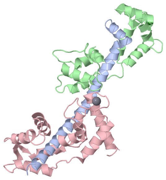
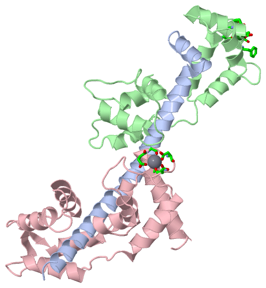
Chain A   (MYS_ARGIR | P24733)
molecular function
    GO:0005524    ATP binding    Interacting selectively and non-covalently with ATP, adenosine 5'-triphosphate, a universally important coenzyme and enzyme regulator.
    GO:0003779    actin binding    Interacting selectively and non-covalently with monomeric or multimeric forms of actin, including actin filaments.
    GO:0005516    calmodulin binding    Interacting selectively and non-covalently with calmodulin, a calcium-binding protein with many roles, both in the calcium-bound and calcium-free states.
    GO:0003774    motor activity    Catalysis of the generation of force resulting either in movement along a microfilament or microtubule, or in torque resulting in membrane scission, coupled to the hydrolysis of a nucleoside triphosphate.
    GO:0000166    nucleotide binding    Interacting selectively and non-covalently with a nucleotide, any compound consisting of a nucleoside that is esterified with (ortho)phosphate or an oligophosphate at any hydroxyl group on the ribose or deoxyribose.
cellular component
    GO:0005737    cytoplasm    All of the contents of a cell excluding the plasma membrane and nucleus, but including other subcellular structures.
    GO:0030016    myofibril    The contractile element of skeletal and cardiac muscle; a long, highly organized bundle of actin, myosin, and other proteins that contracts by a sliding filament mechanism.
    GO:0016459    myosin complex    A protein complex, formed of one or more myosin heavy chains plus associated light chains and other proteins, that functions as a molecular motor; uses the energy of ATP hydrolysis to move actin filaments or to move vesicles or other cargo on fixed actin filaments; has magnesium-ATPase activity and binds actin. Myosin classes are distinguished based on sequence features of the motor, or head, domain, but also have distinct tail regions that are believed to bind specific cargoes.
    GO:0032982    myosin filament    A protein complex containing myosin heavy chains, plus associated light chains and other proteins, in which the myosin heavy chains are arranged into a filament.

Chain B   (MLR_ARGIR | P13543)
molecular function
    GO:0005509    calcium ion binding    Interacting selectively and non-covalently with calcium ions (Ca2+).
    GO:0046872    metal ion binding    Interacting selectively and non-covalently with any metal ion.
cellular component
    GO:0016459    myosin complex    A protein complex, formed of one or more myosin heavy chains plus associated light chains and other proteins, that functions as a molecular motor; uses the energy of ATP hydrolysis to move actin filaments or to move vesicles or other cargo on fixed actin filaments; has magnesium-ATPase activity and binds actin. Myosin classes are distinguished based on sequence features of the motor, or head, domain, but also have distinct tail regions that are believed to bind specific cargoes.

Chain C   (MLE_ARGIR | P07291)
molecular function
    GO:0005509    calcium ion binding    Interacting selectively and non-covalently with calcium ions (Ca2+).
cellular component
    GO:0016459    myosin complex    A protein complex, formed of one or more myosin heavy chains plus associated light chains and other proteins, that functions as a molecular motor; uses the energy of ATP hydrolysis to move actin filaments or to move vesicles or other cargo on fixed actin filaments; has magnesium-ATPase activity and binds actin. Myosin classes are distinguished based on sequence features of the motor, or head, domain, but also have distinct tail regions that are believed to bind specific cargoes.

 Visualization

(-) Interactive Views

Asymmetric/Biological Unit
  Complete Structure
    Jena3D(integrated viewing of ligand, site, SAP, PROSITE, SCOP information)
    WebMol | AstexViewer[tm]@PDBe
(Java Applets, require no local installation except for Java; loading may be slow)
    STRAP
(Java WebStart application, automatic local installation, requires Java; full application with system access!)
    RasMol
(require local installation)
    Molscript (VRML)
(requires installation of a VRML viewer; select preferred view via VRML and generate a mono or stereo PDF format file)
 
  Ligands, Modified Residues, Ions
    CA  [ RasMol | Jena3D ]  +environment [ RasMol | Jena3D ]
    MG  [ RasMol | Jena3D ]  +environment [ RasMol | Jena3D ]
 
  Sites
    AC1  [ RasMol ]  +environment [ RasMol ]
    AC2  [ RasMol ]  +environment [ RasMol ]
 
  Cis Peptide Bonds
(no "Cis Peptide Bonds" information available for 1wdc)
 

(-) Still Images

Jmol
  protein: cartoon or spacefill or dots and stick; nucleic acid: cartoon and stick; ligands: spacefill; active site: stick
Molscript
  protein, nucleic acid: cartoon; ligands: spacefill; active site: ball and stick

 Databases and Analysis Tools

(-) Databases

Access by PDB/NDB ID
  1wdc
    Family and Domain InformationProDom | SYSTERS
    General Structural InformationGlycoscienceDB | MMDB | NDB | OCA | PDB | PDBe | PDBj | PDBsum | PDBWiki | PQS | PROTEOPEDIA
    Orientation in MembranesOPM
    Protein SurfaceSURFACE
    Secondary StructureDSSP (structure derived) | HSSP (homology derived)
    Structural GenomicsGeneCensus
    Structural NeighboursCE | VAST
    Structure ClassificationCATH | Dali | SCOP
    Validation and Original DataBMRB Data View | BMRB Restraints Grid | EDS | PROCHECK | RECOORD | WHAT_CHECK
 
Access by UniProt ID/Accession number
  MLE_ARGIR | P07291
    Comparative Protein Structure ModelsModBase
    Genomic InformationEnsembl
    Protein-protein InteractionDIP
    Sequence, Family and Domain InformationInterPro | Pfam | SMART | UniProtKB/SwissProt
  MLR_ARGIR | P13543
    Comparative Protein Structure ModelsModBase
    Genomic InformationEnsembl
    Protein-protein InteractionDIP
    Sequence, Family and Domain InformationInterPro | Pfam | SMART | UniProtKB/SwissProt
  MYS_ARGIR | P24733
    Comparative Protein Structure ModelsModBase
    Genomic InformationEnsembl
    Protein-protein InteractionDIP
    Sequence, Family and Domain InformationInterPro | Pfam | SMART | UniProtKB/SwissProt
 
Access by Enzyme Classificator   (EC Number)
  (no 'Enzyme Classificator' available)
    General Enzyme InformationBRENDA | EC-PDB | Enzyme | IntEnz
    PathwayKEGG | MetaCyc
 
Access by Disease Identifier   (MIM ID)
  (no 'MIM ID' available)
    Disease InformationOMIM
 
Access by GenAge ID
  (no 'GenAge ID' available)
    Age Related InformationGenAge

(-) Analysis Tools

Access by PDB/NDB ID
    Domain InformationXDom
    Interatomic Contacts of Structural UnitsCSU
    Ligand-protein ContactsLPC
    Protein CavitiescastP
    Sequence and Secondary StructurePDBCartoon
    Structure AlignmentSTRAP(Java WebStart application, automatic local installation, requires Java; full application with system access!)
    Structure and Sequence BrowserSTING
 
Access by UniProt ID/Accession number
  MLE_ARGIR | P07291
    Protein Disorder PredictionDisEMBL | FoldIndex | GLOBPLOT (for more information see DisProt)
  MLR_ARGIR | P13543
    Protein Disorder PredictionDisEMBL | FoldIndex | GLOBPLOT (for more information see DisProt)
  MYS_ARGIR | P24733
    Protein Disorder PredictionDisEMBL | FoldIndex | GLOBPLOT (for more information see DisProt)

 Related Entries

(-) Entries Sharing at Least One Protein Chain (UniProt ID)

UniProtKB/Swiss-Prot
        MLE_ARGIR | P072911b7t 1dfk 1dfl 1kk7 1kk8 1kqm 1kwo 1l2o 1qvi 1s5g 1scm 1sr6 2w4t 2w4v 2w4w 3jtd 3jvt
        MLR_ARGIR | P135431b7t 1dfk 1dfl 1kk7 1kk8 1kqm 1kwo 1l2o 1qvi 1s5g 1scm 1sr6 2w4t 2w4v 2w4w 3jtd 3jvt
        MYS_ARGIR | P247331b7t 1dfk 1dfl 1kk7 1kk8 1kqm 1kwo 1l2o 1qvi 1s5g 1scm 1sr6 2w4t 2w4v 2w4w 3bas 3bat 3jtd 3jvt

(-) Related Entries Specified in the PDB File

(no "Related Entries Specified in the PDB File" available for 1WDC)