Show PDB file:   
         Plain Text   HTML   (compressed file size)
QuickSearch:   
by PDB,NDB,UniProt,PROSITE Code or Search Term(s)  
(-)Asymmetric Unit
(-)Asym. Unit - sites
(-)Biological Unit 1
(-)Biol. Unit 1 - sites
collapse expand < >
Image Asymmetric Unit
Asymmetric Unit  (Jmol Viewer)
Image Asym. Unit - sites
Asym. Unit - sites  (Jmol Viewer)
Image Biological Unit 1
Biological Unit 1  (Jmol Viewer)
Image Biol. Unit 1 - sites
Biol. Unit 1 - sites  (Jmol Viewer)

(-) Description

Title :  KINASE RECRUITMENT DOMAIN OF THE STRESS PHOSPHATASE RSBU
 
Authors :  O. Delumeau, S. Dutta, M. Brigulla, G. Kuhnke, S. W. Hardwick, U. Voelker, M. D. Yudkin, R. J. Lewis
Date :  05 Aug 04  (Deposition) - 05 Aug 04  (Release) - 25 Mar 15  (Revision)
Method :  X-RAY DIFFRACTION
Resolution :  1.60
Chains :  Asym. Unit :  A
Biol. Unit 1:  A  (2x)
Keywords :  Phosphatase, Stress, Bacillus, Kinase, Hydrolase (Keyword Search: [Gene Ontology, PubMed, Web (Google))
 
Reference :  O. Delumeau, S. Dutta, M. Brigulla, G. Kuhnke, S. W. Hardwicku, U. Voelker, M. D. Yudkin, R. J. Lewis
Functional And Structural Characterization Of Rsbu, A Stress Signaling Protein Phosphatase 2C
J. Biol. Chem. V. 279 40927 2004
PubMed-ID: 15263010  |  Reference-DOI: 10.1074/JBC.M405464200

(-) Compounds

Molecule 1 - PHOSPHOSERINE PHOSPHATASE RSBU
    ChainsA
    EngineeredYES
    Expression SystemESCHERICHIA COLI
    Expression System PlasmidPET22B
    Expression System StrainBL21(DE3)
    Expression System Taxid469008
    FragmentN-TERMINAL DOMAIN, RESIDUES 1-84
    Organism ScientificBACILLUS SUBTILIS
    Organism Taxid1423
    SynonymRSBU, SIGMA FACTOR SIGB REGULATION PROTEIN

 Structural Features

(-) Chains, Units

  1
Asymmetric Unit A
Biological Unit 1 (2x)A

Summary Information (see also Sequences/Alignments below)
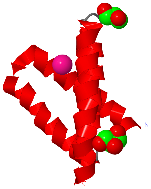
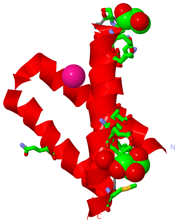
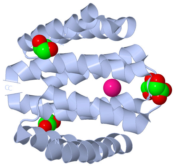
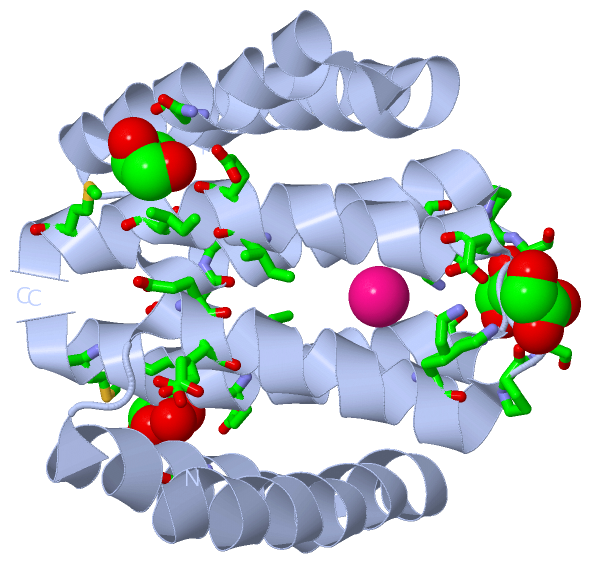
(-) Ligands, Modified Residues, Ions  (2, 3)

Asymmetric Unit (2, 3)
No.NameCountTypeFull Name
1GOL2Ligand/IonGLYCEROL
2XE1Ligand/IonXENON
Biological Unit 1 (1, 4)
No.NameCountTypeFull Name
1GOL4Ligand/IonGLYCEROL
2XE-1Ligand/IonXENON

(-) Sites  (2, 2)

Asymmetric Unit (2, 2)
No.NameEvidenceResiduesDescription
1AC1SOFTWAREGLN A:31 , PRO A:60 , GLU A:75 , ILE A:78 , MET A:82 , HOH A:2034BINDING SITE FOR RESIDUE GOL A1085
2AC2SOFTWAREGLU A:45 , GLU A:46 , SER A:49 , LYS A:56 , PRO A:60 , SER A:61 , GLU A:64 , ILE A:74BINDING SITE FOR RESIDUE GOL A1086

(-) SS Bonds  (0, 0)

(no "SS Bond" information available for 1W53)

(-) Cis Peptide Bonds  (0, 0)

(no "Cis Peptide Bond" information available for 1W53)

 Sequence-Structure Mapping

(-) SAPs(SNPs)/Variants  (0, 0)

(no "SAP(SNP)/Variant" information available for 1W53)

(-) PROSITE Motifs  (0, 0)

(no "PROSITE Motif" information available for 1W53)

(-) Exons   (0, 0)

(no "Exon" information available for 1W53)

(-) Sequences/Alignments

Asymmetric Unit
   Reformat: Number of residues per line =  ('0' or empty: single-line sequence representation)
  Number of residues per labelling interval =   
  UniProt sequence: complete  aligned part    
   Show mapping: SCOP domains CATH domains Pfam domains Secondary structure (by author)
SAPs(SNPs) PROSITE motifs Exons
(details for a mapped element are shown in a popup box when the mouse pointer rests over it)
Chain A from PDB  Type:PROTEIN  Length:84
 aligned with RSBU_BACSU | P40399 from UniProtKB/Swiss-Prot  Length:335

    Alignment length:84
                                    10        20        30        40        50        60        70        80    
            RSBU_BACSU    1 MDFREVIEQRYHQLLSRYIAELTETSLYQAQKFSRKTIEHQIPPEEIISIHRKVLKELYPSLPEDVFHSLDFLIEVMIGYGMAY 84
               SCOP domains d1w53a_ A: Phosphoserine phosphatase RsbU, N-terminal domain                         SCOP domains
               CATH domains 1w53A00 A:1-84 KaiA/RbsU domain                                                      CATH domains
               Pfam domains -------RsbU_N-1w53A01 A:8-84                                                         Pfam domains
         Sec.struct. author hhhhhhhhhhhhhhhhhhhhhhhhhhhhhhhhhhhhhhhh..hhhhhhhhhhhhhhhhh...hhhhhhhhhhhhhhhhhhhhhh Sec.struct. author
                 SAPs(SNPs) ------------------------------------------------------------------------------------ SAPs(SNPs)
                    PROSITE ------------------------------------------------------------------------------------ PROSITE
                 Transcript ------------------------------------------------------------------------------------ Transcript
                  1w53 A  1 MDFREVIEQRYHQLLSRYIAELTETSLYQAQKFSRKTIEHQIPPEEIISIHRKVLKELYPSLPEDVFHSLDFLIEVMIGYGMAY 84
                                    10        20        30        40        50        60        70        80    

   Legend:   → Mismatch (orange background)
  - → Gap (green background, '-', border residues have a numbering label)
    → Modified Residue (blue background, lower-case, 'x' indicates undefined single-letter code, labelled with number + name)
  x → Chemical Group (purple background, 'x', labelled with number + name, e.g. ACE or NH2)
  extra numbering lines below/above indicate numbering irregularities and modified residue names etc., number ends below/above '|'

 Classification and Annotation

(-) SCOP Domains  (1, 1)

Asymmetric Unit

(-) CATH Domains  (1, 1)

Asymmetric Unit

(-) Pfam Domains  (1, 1)

Asymmetric Unit

(-) Gene Ontology  (7, 7)

Asymmetric Unit(hide GO term definitions)
Chain A   (RSBU_BACSU | P40399)
molecular function
    GO:0003824    catalytic activity    Catalysis of a biochemical reaction at physiological temperatures. In biologically catalyzed reactions, the reactants are known as substrates, and the catalysts are naturally occurring macromolecular substances known as enzymes. Enzymes possess specific binding sites for substrates, and are usually composed wholly or largely of protein, but RNA that has catalytic activity (ribozyme) is often also regarded as enzymatic.
    GO:0016787    hydrolase activity    Catalysis of the hydrolysis of various bonds, e.g. C-O, C-N, C-C, phosphoric anhydride bonds, etc. Hydrolase is the systematic name for any enzyme of EC class 3.
    GO:0004721    phosphoprotein phosphatase activity    Catalysis of the reaction: a phosphoprotein + H2O = a protein + phosphate. Together with protein kinases, these enzymes control the state of phosphorylation of cell proteins and thereby provide an important mechanism for regulating cellular activity.
    GO:0004647    phosphoserine phosphatase activity    Catalysis of the reaction: L(or D)-O-phosphoserine + H2O = L(or D)-serine + phosphate.
biological process
    GO:0016311    dephosphorylation    The process of removing one or more phosphoric (ester or anhydride) residues from a molecule.
    GO:0006470    protein dephosphorylation    The process of removing one or more phosphoric residues from a protein.
    GO:0006468    protein phosphorylation    The process of introducing a phosphate group on to a protein.

 Visualization

(-) Interactive Views

Asymmetric Unit
  Complete Structure
    Jena3D(integrated viewing of ligand, site, SAP, PROSITE, SCOP information)
    WebMol | AstexViewer[tm]@PDBe
(Java Applets, require no local installation except for Java; loading may be slow)
    STRAP
(Java WebStart application, automatic local installation, requires Java; full application with system access!)
    RasMol
(require local installation)
    Molscript (VRML)
(requires installation of a VRML viewer; select preferred view via VRML and generate a mono or stereo PDF format file)
 
  Ligands, Modified Residues, Ions
    GOL  [ RasMol | Jena3D ]  +environment [ RasMol | Jena3D ]
    XE  [ RasMol | Jena3D ]  +environment [ RasMol | Jena3D ]
 
  Sites
    AC1  [ RasMol ]  +environment [ RasMol ]
    AC2  [ RasMol ]  +environment [ RasMol ]
 
  Cis Peptide Bonds
(no "Cis Peptide Bonds" information available for 1w53)
 
Biological Unit
  Complete Structure
    Biological Unit 1  [ Jena3D ]

(-) Still Images

Jmol
  protein: cartoon or spacefill or dots and stick; nucleic acid: cartoon and stick; ligands: spacefill; active site: stick
Molscript
  protein, nucleic acid: cartoon; ligands: spacefill; active site: ball and stick

 Databases and Analysis Tools

(-) Databases

Access by PDB/NDB ID
  1w53
    Family and Domain InformationProDom | SYSTERS
    General Structural InformationGlycoscienceDB | MMDB | NDB | OCA | PDB | PDBe | PDBj | PDBsum | PDBWiki | PQS | PROTEOPEDIA
    Orientation in MembranesOPM
    Protein SurfaceSURFACE
    Secondary StructureDSSP (structure derived) | HSSP (homology derived)
    Structural GenomicsGeneCensus
    Structural NeighboursCE | VAST
    Structure ClassificationCATH | Dali | SCOP
    Validation and Original DataBMRB Data View | BMRB Restraints Grid | EDS | PROCHECK | RECOORD | WHAT_CHECK
 
Access by UniProt ID/Accession number
  RSBU_BACSU | P40399
    Comparative Protein Structure ModelsModBase
    Genomic InformationEnsembl
    Protein-protein InteractionDIP
    Sequence, Family and Domain InformationInterPro | Pfam | SMART | UniProtKB/SwissProt
 
Access by Enzyme Classificator   (EC Number)
  (no 'Enzyme Classificator' available)
    General Enzyme InformationBRENDA | EC-PDB | Enzyme | IntEnz
    PathwayKEGG | MetaCyc
 
Access by Disease Identifier   (MIM ID)
  (no 'MIM ID' available)
    Disease InformationOMIM
 
Access by GenAge ID
  (no 'GenAge ID' available)
    Age Related InformationGenAge

(-) Analysis Tools

Access by PDB/NDB ID
    Domain InformationXDom
    Interatomic Contacts of Structural UnitsCSU
    Ligand-protein ContactsLPC
    Protein CavitiescastP
    Sequence and Secondary StructurePDBCartoon
    Structure AlignmentSTRAP(Java WebStart application, automatic local installation, requires Java; full application with system access!)
    Structure and Sequence BrowserSTING
 
Access by UniProt ID/Accession number
  RSBU_BACSU | P40399
    Protein Disorder PredictionDisEMBL | FoldIndex | GLOBPLOT (for more information see DisProt)

 Related Entries

(-) Entries Sharing at Least One Protein Chain (UniProt ID)

UniProtKB/Swiss-Prot
        RSBU_BACSU | P403992j6y 2j6z 2j70

(-) Related Entries Specified in the PDB File

(no "Related Entries Specified in the PDB File" available for 1W53)