|
|
|
|
 Description
|
|
Compounds
|
||||||||||||||||||||||||||||||||||||||||
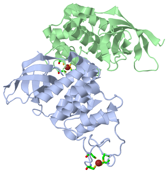
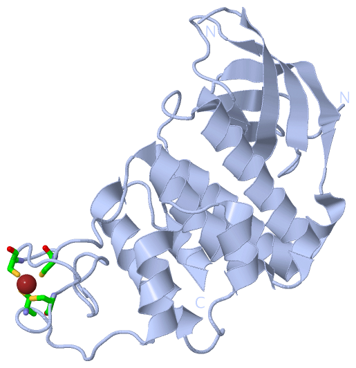
Chains, Units
Summary Information (see also Sequences/Alignments below) |
Ligands, Modified Residues, Ions (1, 2)
Asymmetric Unit (1, 2)
|
Sites (2, 2)
Asymmetric Unit (2, 2)
|
SS Bonds (0, 0)| (no "SS Bond" information available for 1W3S) |
Cis Peptide Bonds (1, 1)
Asymmetric Unit
|
||||||||
SAPs(SNPs)/Variants (0, 0)| (no "SAP(SNP)/Variant" information available for 1W3S) |
PROSITE Motifs (0, 0)| (no "PROSITE Motif" information available for 1W3S) |
Exons (0, 0)| (no "Exon" information available for 1W3S) |
Sequences/Alignments
Asymmetric UnitChain A from PDB Type:PROTEIN Length:230 aligned with Q9RW50_DEIRA | Q9RW50 from UniProtKB/TrEMBL Length:244 Alignment length:236 11 21 31 41 51 61 71 81 91 101 111 121 131 141 151 161 171 181 191 201 211 221 231 Q9RW50_DEIRA 2 RSRTANRSGIVIRRRVTPAGDIIVTLLTPQGKLKAIARGGVKGPLSSSLNLFHHVGVQVYQGPHNDLASVKQAVLEGALPTLAEPERYAFAHLMAEFADALFQEGEFSEQAFDLFAASLRGVAHQPDPEWVALVMSYKLLGLAGVIPQTARCARCGAPDPEHPDPLGGQLLCSKCAALPPYPPAVLDFLRHAVRRTVRASFEQPVPSADRPALWRALEKFVTVQVGGVHSWRQLVP 237 SCOP domains d1w3sa1 A:2-80 Recombinational repair p rotein RecO, N-terminal domain d1w3sa2 A:81-237 Recombinational repair protein RecO, C-terminal domain SCOP domains CATH domains -------------------------------------------------------------------------------------------------------------------------------------------------------------------------------------------------------------------------------------------- CATH domains Pfam domains -------------------------------------------------------------------------------------------------------------------------------------------------------------------------------------------------------------------------------------------- Pfam domains SAPs(SNPs) -------------------------------------------------------------------------------------------------------------------------------------------------------------------------------------------------------------------------------------------- SAPs(SNPs) PROSITE -------------------------------------------------------------------------------------------------------------------------------------------------------------------------------------------------------------------------------------------- PROSITE Transcript -------------------------------------------------------------------------------------------------------------------------------------------------------------------------------------------------------------------------------------------- Transcript 1w3s A 2 RSRTANRSGIVIRRRVTPAGDIIVTLLTPQGKLKAIARG------SSSLNLFHHVGVQVYQGPHNDLASVKQAVLEGALPTLAEPERYAFAHLMAEFADALFQEGEFSEQAFDLFAASLRGVAHQPDPEWVALVMSYKLLGLAGVIPQTARCARCGAPDPEHPDPLGGQLLCSKCAALPPYPPAVLDFLRHAVRRTVRASFEQPVPSADRPALWRALEKFVTVQVGGVHSWRQLVP 237 11 21 31 |- | 51 61 71 81 91 101 111 121 131 141 151 161 171 181 191 201 211 221 231 40 47 Chain B from PDB Type:PROTEIN Length:231 aligned with Q9RW50_DEIRA | Q9RW50 from UniProtKB/TrEMBL Length:244 Alignment length:236 12 22 32 42 52 62 72 82 92 102 112 122 132 142 152 162 172 182 192 202 212 222 232 Q9RW50_DEIRA 3 SRTANRSGIVIRRRVTPAGDIIVTLLTPQGKLKAIARGGVKGPLSSSLNLFHHVGVQVYQGPHNDLASVKQAVLEGALPTLAEPERYAFAHLMAEFADALFQEGEFSEQAFDLFAASLRGVAHQPDPEWVALVMSYKLLGLAGVIPQTARCARCGAPDPEHPDPLGGQLLCSKCAALPPYPPAVLDFLRHAVRRTVRASFEQPVPSADRPALWRALEKFVTVQVGGVHSWRQLVPS 238 SCOP domains d1w3sb1 B:3-80 Recombinational repair protein RecO, N-terminal domain d1w3sb2 B:81-238 Recombinational repair protein RecO, C-terminal domain SCOP domains CATH domains -------------------------------------------------------------------------------------------------------------------------------------------------------------------------------------------------------------------------------------------- CATH domains Pfam domains (1) RecO_N-1w3sB01 B:3-79 ---RecO_C-1w3sB03 B:83-233 ----- Pfam domains (1) Pfam domains (2) RecO_N-1w3sB02 B:3-79 ---RecO_C-1w3sB04 B:83-233 ----- Pfam domains (2) SAPs(SNPs) -------------------------------------------------------------------------------------------------------------------------------------------------------------------------------------------------------------------------------------------- SAPs(SNPs) PROSITE -------------------------------------------------------------------------------------------------------------------------------------------------------------------------------------------------------------------------------------------- PROSITE Transcript -------------------------------------------------------------------------------------------------------------------------------------------------------------------------------------------------------------------------------------------- Transcript 1w3s B 3 SRTANRSGIVIRRRVTPAGDIIVTLLTPQGKLKAIARG-----LSSSLNLFHHVGVQVYQGPHNDLASVKQAVLEGALPTLAEPERYAFAHLMAEFADALFQEGEFSEQAFDLFAASLRGVAHQPDPEWVALVMSYKLLGLAGVIPQTARCARCGAPDPEHPDPLGGQLLCSKCAALPPYPPAVLDFLRHAVRRTVRASFEQPVPSADRPALWRALEKFVTVQVGGVHSWRQLVPS 238 12 22 32 | - | 52 62 72 82 92 102 112 122 132 142 152 162 172 182 192 202 212 222 232 40 46
|
||||||||||||||||||||
SCOP Domains (2, 4)
Asymmetric Unit
 |
CATH Domains (0, 0)| (no "CATH Domain" information available for 1W3S) |
Pfam Domains (2, 4)
Asymmetric Unit
|
Gene Ontology (5, 5)|
Asymmetric Unit(hide GO term definitions) Chain A,B (Q9RW50_DEIRA | Q9RW50)
|
||||||||||||||||||||||||||||||||||||||||||
Interactive Views
|
|||||||||||||||||||||||||||||||||||||||||||||||||||||||||||||||||||||||||||||||||||||||||||||||||||||||||||||||||||||||||||||||||||||||||||||||||||||
Still Images
|
||||||||||||||||
Databases
|
||||||||||||||||||||||||||||||||||||||||||||||||||||||||||||||||||||||||||||||||||||||||||||||||||||||||||||||||||||||||||||||||||||||||||||||||||||||||||||||||
Analysis Tools
|
|||||||||||||||||||||||||||||||||||||||||||||||||||||||||||||
Entries Sharing at Least One Protein Chain (UniProt ID)
Related Entries Specified in the PDB File
|
|