Show PDB file:   
         Plain Text   HTML   (compressed file size)
QuickSearch:   
by PDB,NDB,UniProt,PROSITE Code or Search Term(s)  
(-)Asymmetric Unit
(-)Biological Unit 1
(-)Biological Unit 2
collapse expand < >
Image Asymmetric Unit
Asymmetric Unit  (Jmol Viewer)
Image Biological Unit 1
Biological Unit 1  (Jmol Viewer)
Image Biological Unit 2
Biological Unit 2  (Jmol Viewer)

(-) Description

Title :  STRUCTURE OF BACILLUS SUBTILIS PDAA, A FAMILY 4 CARBOHYDRATE ESTERASE.
 
Authors :  D. E. Blair, D. M. F. Van Aalten
Date :  17 Jun 04  (Deposition) - 10 Jan 05  (Release) - 24 Feb 09  (Revision)
Method :  X-RAY DIFFRACTION
Resolution :  1.90
Chains :  Asym. Unit :  A,B
Biol. Unit 1:  A  (1x)
Biol. Unit 2:  B  (1x)
Keywords :  Hydrolase, Family 4 Carbohydrate Esterase, Deacetylase, Peptidoglycan, Nodb Homology Domain (Keyword Search: [Gene Ontology, PubMed, Web (Google))
 
Reference :  D. E. Blair, D. M. F. Van Aalten
Structure Of Bacillus Subtilis Pdaa, A Family 4 Carbohydrate Esterase, And A Complex With N-Acetyl- Glucosamine
Febs Lett. V. 570 13 2004
PubMed-ID: 15251431  |  Reference-DOI: 10.1016/J.FEBSLET.2004.06.013

(-) Compounds

Molecule 1 - PROBABLE POLYSACCHARIDE DEACETYLASE PDAA
    ChainsA, B
    EC Number3.-.-.-
    EngineeredYES
    Expression SystemESCHERICHIA COLI
    Expression System Cell LineBL21DE3
    Expression System PlasmidPGEX-6P-1
    Expression System Taxid562
    Organism ScientificBACILLUS SUBTILIS
    Organism Taxid1423
    SynonymPDAA

 Structural Features

(-) Chains, Units

  12
Asymmetric Unit AB
Biological Unit 1 (1x)A 
Biological Unit 2 (1x) B

Summary Information (see also Sequences/Alignments below)

(-) Ligands, Modified Residues, Ions  (0, 0)

(no "Ligand,Modified Residues,Ions" information available for 1W17)

(-) Sites  (0, 0)

(no "Site" information available for 1W17)

(-) SS Bonds  (0, 0)

(no "SS Bond" information available for 1W17)

(-) Cis Peptide Bonds  (1, 1)
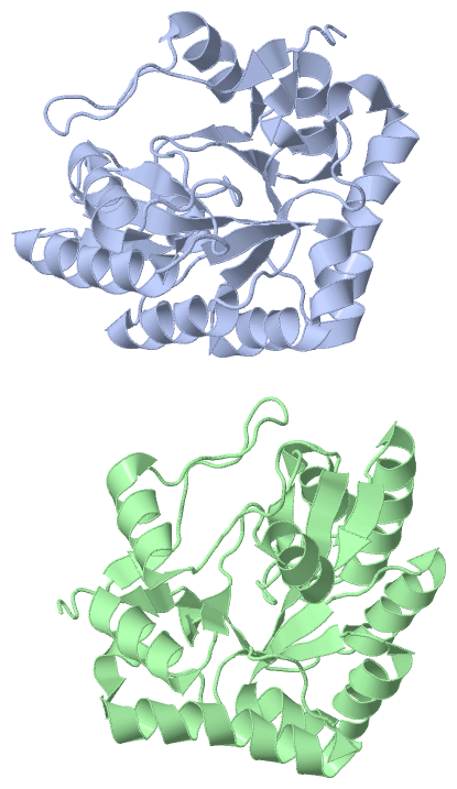
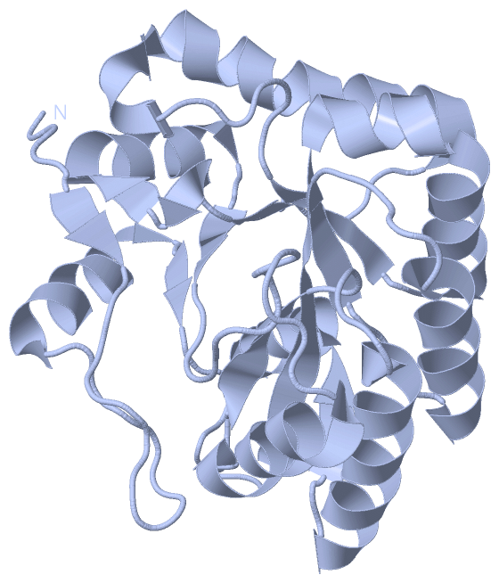
Asymmetric Unit
No.Residues
1Val A:24 -Pro A:25

 Sequence-Structure Mapping

(-) SAPs(SNPs)/Variants  (0, 0)

(no "SAP(SNP)/Variant" information available for 1W17)

(-) PROSITE Motifs  (1, 2)

Asymmetric Unit (1, 2)
 PROSITEUniProtKBPDB
No.IDACDescriptionIDLocationCountLocation
1NODBPS51677 NodB homology domain profile.PDAA_BACSU66-247
 
  2A:66-247
B:66-247
Biological Unit 1 (1, 1)
 PROSITEUniProtKBPDB
No.IDACDescriptionIDLocationCountLocation
1NODBPS51677 NodB homology domain profile.PDAA_BACSU66-247
 
  1A:66-247
-
Biological Unit 2 (1, 1)
 PROSITEUniProtKBPDB
No.IDACDescriptionIDLocationCountLocation
1NODBPS51677 NodB homology domain profile.PDAA_BACSU66-247
 
  1-
B:66-247

(-) Exons   (0, 0)

(no "Exon" information available for 1W17)

(-) Sequences/Alignments

Asymmetric Unit
   Reformat: Number of residues per line =  ('0' or empty: single-line sequence representation)
  Number of residues per labelling interval =   
  UniProt sequence: complete  aligned part    
   Show mapping: SCOP domains CATH domains Pfam domains Secondary structure (by author)
SAPs(SNPs) PROSITE motifs Exons
(details for a mapped element are shown in a popup box when the mouse pointer rests over it)
Chain A from PDB  Type:PROTEIN  Length:236
 aligned with PDAA_BACSU | O34928 from UniProtKB/Swiss-Prot  Length:263

    Alignment length:236
                                    33        43        53        63        73        83        93       103       113       123       133       143       153       163       173       183       193       203       213       223       233       243       253      
           PDAA_BACSU    24 VPNEPINWGFKRSVNHQPPDAGKQLNSLIEKYDAFYLGNTKEKTIYLTFDNGYENGYTPKVLDVLKKHRVTGTFFVTGHFVKDQPQLIKRMSDEGHIIGNHSFHHPDLTTKTADQIQDELDSVNEEVYKITGKQDNLYLRPPRGVFSEYVLKETKRLGYQTVFWSVAFVDWKINNQKGKKYAYDHMIKQAHPGAIYLLHTVSRDNAEALDDAITDLKKQGYTFKSIDDLMFEKEMR 259
               SCOP domains d1w17a_ A: automated matches                                                                                                                                                                                                                 SCOP domains
               CATH domains 1w17A00 A:24-259 Glycoside hydrolase/deacetylase                                                                                                                                                                                             CATH domains
               Pfam domains -------------------------------------------------------------------------------------------------------------------------------------------------------------------------------------------------------------------------------------------- Pfam domains
         Sec.struct. author .....ee..............hhhhhhhhhhh.ee........eeeeeeee.....hhhhhhhhhhhh....eeeehhhhhhhhhhhhhhhhhh..eeee......hhhhhhhhhhhhhhhhhhhhhhhhhh.....ee.hhh.eehhhhhhhhhhh..eee...............hhhhhhhhhhhh....eeeee.....hhhhhhhhhhhhhhhh..eeehhhhhhhhhhh. Sec.struct. author
                 SAPs(SNPs) -------------------------------------------------------------------------------------------------------------------------------------------------------------------------------------------------------------------------------------------- SAPs(SNPs)
                    PROSITE ------------------------------------------NODB  PDB: A:66-247 UniProt: 66-247                                                                                                                                                   ------------ PROSITE
                 Transcript -------------------------------------------------------------------------------------------------------------------------------------------------------------------------------------------------------------------------------------------- Transcript
                 1w17 A  24 VPNEPINWGFKRSVNHQPPDAGKQLNSLIEKYDAFYLGNTKEKTIYLTFDNGYENGYTPKVLDVLKKHRVTGTFFVTGHFVKDQPQLIKRMSDEGHIIGNHSFHHPDLTTKTADQIQDELDSVNEEVYKITGKQDNLYLRPPRGVFSEYVLKETKRLGYQTVFWSVAFVDWKINNQKGKKYAYDHMIKQAHPGAIYLLHTVSRDNAEALDDAITDLKKQGYTFKSIDDLMFEKEMR 259
                                    33        43        53        63        73        83        93       103       113       123       133       143       153       163       173       183       193       203       213       223       233       243       253      

Chain B from PDB  Type:PROTEIN  Length:235
 aligned with PDAA_BACSU | O34928 from UniProtKB/Swiss-Prot  Length:263

    Alignment length:235
                                    33        43        53        63        73        83        93       103       113       123       133       143       153       163       173       183       193       203       213       223       233       243       253     
           PDAA_BACSU    24 VPNEPINWGFKRSVNHQPPDAGKQLNSLIEKYDAFYLGNTKEKTIYLTFDNGYENGYTPKVLDVLKKHRVTGTFFVTGHFVKDQPQLIKRMSDEGHIIGNHSFHHPDLTTKTADQIQDELDSVNEEVYKITGKQDNLYLRPPRGVFSEYVLKETKRLGYQTVFWSVAFVDWKINNQKGKKYAYDHMIKQAHPGAIYLLHTVSRDNAEALDDAITDLKKQGYTFKSIDDLMFEKEM 258
               SCOP domains d1w17b_ B: automated matches                                                                                                                                                                                                                SCOP domains
               CATH domains 1w17B00 B:24-258 Glycoside hydrolase/deacetylase                                                                                                                                                                                            CATH domains
           Pfam domains (1) ------------------------------------Polysacc_deac_1-1w17B01 B:60-185                                                                                              ------------------------------------------------------------------------- Pfam domains (1)
           Pfam domains (2) ------------------------------------Polysacc_deac_1-1w17B02 B:60-185                                                                                              ------------------------------------------------------------------------- Pfam domains (2)
         Sec.struct. author .....ee..............hhhhhhhhhhh...........eeeeeeee.....hhhhhhhhhhhh....eeeehhhhhhhhhhhhhhhhhh..eeee......hhhhhhhhhhhhhhhhhhhhhhhhhh.....ee.hhh.eehhhhhhhhhhh..ee................hhhhhhhhhhhh....eeeee.....hhhhhhhhhhhhhhhh..eeehhhhhhhhhh. Sec.struct. author
                 SAPs(SNPs) ------------------------------------------------------------------------------------------------------------------------------------------------------------------------------------------------------------------------------------------- SAPs(SNPs)
                    PROSITE ------------------------------------------NODB  PDB: B:66-247 UniProt: 66-247                                                                                                                                                   ----------- PROSITE
                 Transcript ------------------------------------------------------------------------------------------------------------------------------------------------------------------------------------------------------------------------------------------- Transcript
                 1w17 B  24 VPNEPINWGFKRSVNHQPPDAGKQLNSLIEKYDAFYLGNTKEKTIYLTFDNGYENGYTPKVLDVLKKHRVTGTFFVTGHFVKDQPQLIKRMSDEGHIIGNHSFHHPDLTTKTADQIQDELDSVNEEVYKITGKQDNLYLRPPRGVFSEYVLKETKRLGYQTVFWSVAFVDWKINNQKGKKYAYDHMIKQAHPGAIYLLHTVSRDNAEALDDAITDLKKQGYTFKSIDDLMFEKEM 258
                                    33        43        53        63        73        83        93       103       113       123       133       143       153       163       173       183       193       203       213       223       233       243       253     

   Legend:   → Mismatch (orange background)
  - → Gap (green background, '-', border residues have a numbering label)
    → Modified Residue (blue background, lower-case, 'x' indicates undefined single-letter code, labelled with number + name)
  x → Chemical Group (purple background, 'x', labelled with number + name, e.g. ACE or NH2)
  extra numbering lines below/above indicate numbering irregularities and modified residue names etc., number ends below/above '|'

 Classification and Annotation

(-) SCOP Domains  (1, 2)

Asymmetric Unit

(-) CATH Domains  (1, 2)

Asymmetric Unit
(-)
Class: Alpha Beta (26913)

(-) Pfam Domains  (1, 2)

Asymmetric Unit
(-)
Clan: GH_CE (28)

(-) Gene Ontology  (7, 7)

Asymmetric Unit(hide GO term definitions)
Chain A,B   (PDAA_BACSU | O34928)
molecular function
    GO:0003824    catalytic activity    Catalysis of a biochemical reaction at physiological temperatures. In biologically catalyzed reactions, the reactants are known as substrates, and the catalysts are naturally occurring macromolecular substances known as enzymes. Enzymes possess specific binding sites for substrates, and are usually composed wholly or largely of protein, but RNA that has catalytic activity (ribozyme) is often also regarded as enzymatic.
    GO:0016787    hydrolase activity    Catalysis of the hydrolysis of various bonds, e.g. C-O, C-N, C-C, phosphoric anhydride bonds, etc. Hydrolase is the systematic name for any enzyme of EC class 3.
    GO:0016810    hydrolase activity, acting on carbon-nitrogen (but not peptide) bonds    Catalysis of the hydrolysis of any carbon-nitrogen bond, C-N, with the exception of peptide bonds.
    GO:0046872    metal ion binding    Interacting selectively and non-covalently with any metal ion.
biological process
    GO:0005975    carbohydrate metabolic process    The chemical reactions and pathways involving carbohydrates, any of a group of organic compounds based of the general formula Cx(H2O)y. Includes the formation of carbohydrate derivatives by the addition of a carbohydrate residue to another molecule.
    GO:0071555    cell wall organization    A process that results in the assembly, arrangement of constituent parts, or disassembly of the cell wall, the rigid or semi-rigid envelope lying outside the cell membrane of plant, fungal and most prokaryotic cells, maintaining their shape and protecting them from osmotic lysis.
    GO:0030435    sporulation resulting in formation of a cellular spore    The process in which a relatively unspecialized cell acquires the specialized features of a cellular spore, a cell form that can be used for dissemination, for survival of adverse conditions because of its heat and dessication resistance, and/or for reproduction.

 Visualization

(-) Interactive Views

Asymmetric Unit
  Complete Structure
    Jena3D(integrated viewing of ligand, site, SAP, PROSITE, SCOP information)
    WebMol | AstexViewer[tm]@PDBe
(Java Applets, require no local installation except for Java; loading may be slow)
    STRAP
(Java WebStart application, automatic local installation, requires Java; full application with system access!)
    RasMol
(require local installation)
    Molscript (VRML)
(requires installation of a VRML viewer; select preferred view via VRML and generate a mono or stereo PDF format file)
 
  Ligands, Modified Residues, Ions
(no "Ligands, Modified Residues, Ions" information available for 1w17)
 
  Sites
(no "Sites" information available for 1w17)
 
  Cis Peptide Bonds
    Val A:24 - Pro A:25   [ RasMol ]  
 
Biological Units
  Complete Structure
    Biological Unit 1  [ Jena3D ]
    Biological Unit 2  [ Jena3D ]

(-) Still Images

Jmol
  protein: cartoon or spacefill or dots and stick; nucleic acid: cartoon and stick; ligands: spacefill; active site: stick
Molscript
  protein, nucleic acid: cartoon; ligands: spacefill; active site: ball and stick

 Databases and Analysis Tools

(-) Databases

Access by PDB/NDB ID
  1w17
    Family and Domain InformationProDom | SYSTERS
    General Structural InformationGlycoscienceDB | MMDB | NDB | OCA | PDB | PDBe | PDBj | PDBsum | PDBWiki | PQS | PROTEOPEDIA
    Orientation in MembranesOPM
    Protein SurfaceSURFACE
    Secondary StructureDSSP (structure derived) | HSSP (homology derived)
    Structural GenomicsGeneCensus
    Structural NeighboursCE | VAST
    Structure ClassificationCATH | Dali | SCOP
    Validation and Original DataBMRB Data View | BMRB Restraints Grid | EDS | PROCHECK | RECOORD | WHAT_CHECK
 
Access by UniProt ID/Accession number
  PDAA_BACSU | O34928
    Comparative Protein Structure ModelsModBase
    Genomic InformationEnsembl
    Protein-protein InteractionDIP
    Sequence, Family and Domain InformationInterPro | Pfam | SMART | UniProtKB/SwissProt
 
Access by Enzyme Classificator   (EC Number)
  3.-.-.-
    General Enzyme InformationBRENDA | EC-PDB | Enzyme | IntEnz
    PathwayKEGG | MetaCyc
 
Access by Disease Identifier   (MIM ID)
  (no 'MIM ID' available)
    Disease InformationOMIM
 
Access by GenAge ID
  (no 'GenAge ID' available)
    Age Related InformationGenAge

(-) Analysis Tools

Access by PDB/NDB ID
    Domain InformationXDom
    Interatomic Contacts of Structural UnitsCSU
    Ligand-protein ContactsLPC
    Protein CavitiescastP
    Sequence and Secondary StructurePDBCartoon
    Structure AlignmentSTRAP(Java WebStart application, automatic local installation, requires Java; full application with system access!)
    Structure and Sequence BrowserSTING
 
Access by UniProt ID/Accession number
  PDAA_BACSU | O34928
    Protein Disorder PredictionDisEMBL | FoldIndex | GLOBPLOT (for more information see DisProt)

 Related Entries

(-) Entries Sharing at Least One Protein Chain (UniProt ID)

UniProtKB/Swiss-Prot
        PDAA_BACSU | O349281ny1 1w1a 1w1b

(-) Related Entries Specified in the PDB File

1ny1 CRYSTAL STRUCTURE OF B. SUBTILIS POLYSACCHARIDE DEACETYLASENORTHEAST STRUCTURAL GENOMICS CONSORTIUM TARGET SR127.
1w1a STRUCTURE OF BACILLUS SUBTILIS PDAA IN COMPLEX WITH NAG, A FAMILY 4 CARBOHYDRATE ESTERASE.
1w1b STRUCTURE OF BACILLUS SUBTILIS PDAA WITH CADMIUM, A FAMILY 4 CARBOHYDRATE ESTERASE.