Show PDB file:   
         Plain Text   HTML   (compressed file size)
QuickSearch:   
by PDB,NDB,UniProt,PROSITE Code or Search Term(s)  
(-)Asym./Biol. Unit
(-)Asym./Biol. Unit - sites
collapse expand < >
Image Asym./Biol. Unit
Asym./Biol. Unit  (Jmol Viewer)
Image Asym./Biol. Unit - sites
Asym./Biol. Unit - sites  (Jmol Viewer)

(-) Description

Title :  CRYSTAL STRUCTURE OF A PUTATIVE HEME OXYGENASE (ALR5027) FROM NOSTOC SP. PCC 7120 AT 1.50 A RESOLUTION
 
Authors :  Joint Center For Structural Genomics (Jcsg)
Date :  13 Jul 04  (Deposition) - 17 Aug 04  (Release) - 13 Jul 11  (Revision)
Method :  X-RAY DIFFRACTION
Resolution :  1.50
Chains :  Asym./Biol. Unit :  A,B
Keywords :  Structural Genomics, Joint Center For Structural Genomics, Jcsg, Protein Structure Initiative, Psi, Oxidoreductase (Keyword Search: [Gene Ontology, PubMed, Web (Google))
 
Reference :  Joint Center For Structural Genomics (Jcsg)
Crystal Structure Of Hypothetical Protein (Alr5027) From Nostoc Sp. At 1. 50 A Resolution
To Be Published
PubMed: search

(-) Compounds

Molecule 1 - HYPOTHETICAL PROTEIN ALR5027
    ChainsA, B
    EngineeredYES
    Expression SystemESCHERICHIA COLI
    Expression System Taxid562
    Expression System Vector TypePLASMID
    GeneALR5027
    Organism ScientificNOSTOC SP.
    Organism Taxid103690
    StrainPCC 7120

 Structural Features

(-) Chains, Units

  12
Asymmetric/Biological Unit AB

Summary Information (see also Sequences/Alignments below)

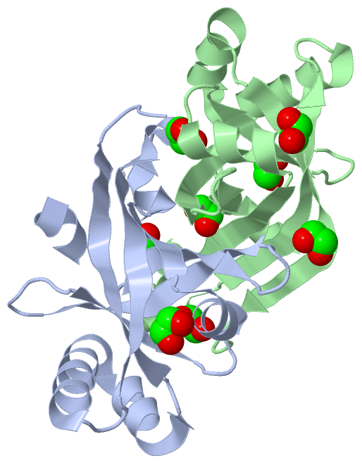
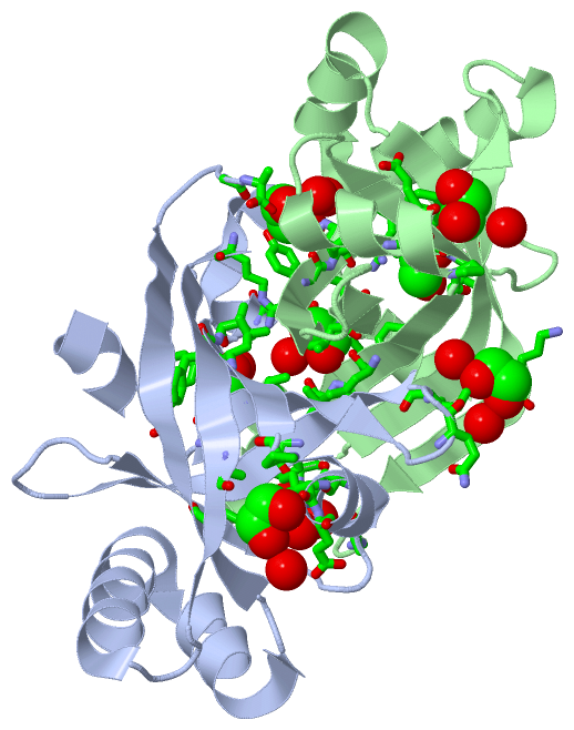
(-) Ligands, Modified Residues, Ions  (1, 8)

Asymmetric/Biological Unit (1, 8)
No.NameCountTypeFull Name
1EDO8Ligand/Ion1,2-ETHANEDIOL

(-) Sites  (8, 8)

Asymmetric Unit (8, 8)
No.NameEvidenceResiduesDescription
1AC1SOFTWARETYR A:36 , HIS A:57 , GLY B:144 , ALA B:145 , HOH B:156 , HOH B:160BINDING SITE FOR RESIDUE EDO B 146
2AC2SOFTWAREGLY A:144 , ALA A:145 , HOH A:152 , HOH A:157 , TYR B:36 , ALA B:37 , HIS B:57BINDING SITE FOR RESIDUE EDO A 146
3AC3SOFTWAREPHE A:18 , SER A:20 , ILE A:22 , LEU A:70 , PHE A:71 , VAL A:72 , HOH A:169 , GLU B:75 , ARG B:84BINDING SITE FOR RESIDUE EDO A 147
4AC4SOFTWAREILE B:61 , GLU B:62 , PRO B:65 , PHE B:130 , HOH B:158BINDING SITE FOR RESIDUE EDO B 147
5AC5SOFTWAREILE A:61 , GLU A:62 , PRO A:65 , ALA A:93 , PHE A:130 , HOH A:226BINDING SITE FOR RESIDUE EDO A 148
6AC6SOFTWAREVAL B:40 , ILE B:41 , ARG B:113 , HOH B:235BINDING SITE FOR RESIDUE EDO B 148
7AC7SOFTWAREGLU A:75 , ARG A:84 , PHE B:18 , SER B:20 , ILE B:22 , LEU B:70 , PHE B:71 , VAL B:72 , HOH B:186BINDING SITE FOR RESIDUE EDO B 149
8AC8SOFTWAREGLN A:29 , ILE A:31 , LYS B:135 , HOH B:157 , HOH B:226BINDING SITE FOR RESIDUE EDO B 150

(-) SS Bonds  (0, 0)

(no "SS Bond" information available for 1VL7)

(-) Cis Peptide Bonds  (0, 0)

(no "Cis Peptide Bond" information available for 1VL7)

 Sequence-Structure Mapping

(-) SAPs(SNPs)/Variants  (0, 0)

(no "SAP(SNP)/Variant" information available for 1VL7)

(-) PROSITE Motifs  (0, 0)

(no "PROSITE Motif" information available for 1VL7)

(-) Exons   (0, 0)

(no "Exon" information available for 1VL7)

(-) Sequences/Alignments

Asymmetric/Biological Unit
   Reformat: Number of residues per line =  ('0' or empty: single-line sequence representation)
  Number of residues per labelling interval =   
  UniProt sequence: complete  aligned part    
   Show mapping: SCOP domains CATH domains Pfam domains Secondary structure (by author)
SAPs(SNPs) PROSITE motifs Exons
(details for a mapped element are shown in a popup box when the mouse pointer rests over it)
Chain A from PDB  Type:PROTEIN  Length:135
 aligned with Q8YMA7_NOSS1 | Q8YMA7 from UniProtKB/TrEMBL  Length:165

    Alignment length:135
                                    20        30        40        50        60        70        80        90       100       110       120       130       140     
         Q8YMA7_NOSS1    11 YAGFIQEFQSAIISTISEQGIPNGSYAPFVIDDAKNIYIYVSGLAVHTKNIEANPLVNVLFVDDEAKTNQIFARRRLSFDCTATLIERESQKWNQVVDQFQERFGQIIEVLRGLADFRIFQLTPKEGRFVIGFGA 145
               SCOP domains d1vl7a_ A: Hypothetical protein Alr5027                                                                                                 SCOP domains
               CATH domains 1vl7A00 A:11-145 Electron Transport, Fmn-binding Protein; Chain A                                                                       CATH domains
               Pfam domains --------------------------------------------------------------------------------------------------------------------------------------- Pfam domains
         Sec.struct. author hhhhhh...eeeeeee.....eeeeeeeeee.....eeeee...hhhhhhhhhh.eeeeeee.hhhhh.hhhhh.eeeeeeeeeee...hhhhhhhhhhhhhhhhhhhhhhhhhh..eeeeeeeeeeeee..... Sec.struct. author
                 SAPs(SNPs) --------------------------------------------------------------------------------------------------------------------------------------- SAPs(SNPs)
                    PROSITE --------------------------------------------------------------------------------------------------------------------------------------- PROSITE
                 Transcript --------------------------------------------------------------------------------------------------------------------------------------- Transcript
                 1vl7 A  11 YAGFIQEFQSAIISTISEQGIPNGSYAPFVIDDAKNIYIYVSGLAVHTKNIEANPLVNVLFVDDEAKTNQIFARRRLSFDCTATLIERESQKWNQVVDQFQERFGQIIEVLRGLADFRIFQLTPKEGRFVIGFGA 145
                                    20        30        40        50        60        70        80        90       100       110       120       130       140     

Chain B from PDB  Type:PROTEIN  Length:135
 aligned with Q8YMA7_NOSS1 | Q8YMA7 from UniProtKB/TrEMBL  Length:165

    Alignment length:135
                                    20        30        40        50        60        70        80        90       100       110       120       130       140     
         Q8YMA7_NOSS1    11 YAGFIQEFQSAIISTISEQGIPNGSYAPFVIDDAKNIYIYVSGLAVHTKNIEANPLVNVLFVDDEAKTNQIFARRRLSFDCTATLIERESQKWNQVVDQFQERFGQIIEVLRGLADFRIFQLTPKEGRFVIGFGA 145
               SCOP domains d1vl7b_ B: Hypothetical protein Alr5027                                                                                                 SCOP domains
               CATH domains 1vl7B00 B:11-145 Electron Transport, Fmn-binding Protein; Chain A                                                                       CATH domains
           Pfam domains (1) Pyridox_oxidase-1vl7B01 B:11-100                                                          --------------------------------------------- Pfam domains (1)
           Pfam domains (2) Pyridox_oxidase-1vl7B02 B:11-100                                                          --------------------------------------------- Pfam domains (2)
         Sec.struct. author hhhhhh...eeeeeee.....eeeeeeeeee.....eeeee...hhhhhhhhhh.eeeeeee.hhhhh.hhhhh.eeeeeeeeeee...hhhhhhhhhhhhhhhhhhhhhhhhhh..eeeeeeeeeeeee..... Sec.struct. author
                 SAPs(SNPs) --------------------------------------------------------------------------------------------------------------------------------------- SAPs(SNPs)
                    PROSITE --------------------------------------------------------------------------------------------------------------------------------------- PROSITE
                 Transcript --------------------------------------------------------------------------------------------------------------------------------------- Transcript
                 1vl7 B  11 YAGFIQEFQSAIISTISEQGIPNGSYAPFVIDDAKNIYIYVSGLAVHTKNIEANPLVNVLFVDDEAKTNQIFARRRLSFDCTATLIERESQKWNQVVDQFQERFGQIIEVLRGLADFRIFQLTPKEGRFVIGFGA 145
                                    20        30        40        50        60        70        80        90       100       110       120       130       140     

   Legend:   → Mismatch (orange background)
  - → Gap (green background, '-', border residues have a numbering label)
    → Modified Residue (blue background, lower-case, 'x' indicates undefined single-letter code, labelled with number + name)
  x → Chemical Group (purple background, 'x', labelled with number + name, e.g. ACE or NH2)
  extra numbering lines below/above indicate numbering irregularities and modified residue names etc., number ends below/above '|'

 Classification and Annotation

(-) SCOP Domains  (1, 2)

Asymmetric/Biological Unit

(-) CATH Domains  (1, 2)

Asymmetric/Biological Unit
(-)
Class: Mainly Beta (13760)

(-) Pfam Domains  (1, 2)

Asymmetric/Biological Unit

(-) Gene Ontology  (5, 5)

Asymmetric/Biological Unit(hide GO term definitions)
Chain A,B   (Q8YMA7_NOSS1 | Q8YMA7)
molecular function
    GO:0010181    FMN binding    Interacting selectively and non-covalently with flavin mono nucleotide. Flavin mono nucleotide (FMN) is the coenzyme or the prosthetic group of various flavoprotein oxidoreductase enzymes.
    GO:0016491    oxidoreductase activity    Catalysis of an oxidation-reduction (redox) reaction, a reversible chemical reaction in which the oxidation state of an atom or atoms within a molecule is altered. One substrate acts as a hydrogen or electron donor and becomes oxidized, while the other acts as hydrogen or electron acceptor and becomes reduced.
    GO:0004733    pyridoxamine-phosphate oxidase activity    Catalysis of the reaction: pyridoxamine 5'-phosphate + H2O + O2 = pyridoxal 5'-phosphate + NH3 + hydrogen peroxide.
biological process
    GO:0055114    oxidation-reduction process    A metabolic process that results in the removal or addition of one or more electrons to or from a substance, with or without the concomitant removal or addition of a proton or protons.
    GO:0042823    pyridoxal phosphate biosynthetic process    The chemical reactions and pathways resulting in the formation of pyridoxal phosphate, pyridoxal phosphorylated at the hydroxymethyl group of C-5, the active form of vitamin B6.

 Visualization

(-) Interactive Views

Asymmetric/Biological Unit
  Complete Structure
    Jena3D(integrated viewing of ligand, site, SAP, PROSITE, SCOP information)
    WebMol | AstexViewer[tm]@PDBe
(Java Applets, require no local installation except for Java; loading may be slow)
    STRAP
(Java WebStart application, automatic local installation, requires Java; full application with system access!)
    RasMol
(require local installation)
    Molscript (VRML)
(requires installation of a VRML viewer; select preferred view via VRML and generate a mono or stereo PDF format file)
 
  Ligands, Modified Residues, Ions
    EDO  [ RasMol | Jena3D ]  +environment [ RasMol | Jena3D ]
 
  Sites
    AC1  [ RasMol ]  +environment [ RasMol ]
    AC2  [ RasMol ]  +environment [ RasMol ]
    AC3  [ RasMol ]  +environment [ RasMol ]
    AC4  [ RasMol ]  +environment [ RasMol ]
    AC5  [ RasMol ]  +environment [ RasMol ]
    AC6  [ RasMol ]  +environment [ RasMol ]
    AC7  [ RasMol ]  +environment [ RasMol ]
    AC8  [ RasMol ]  +environment [ RasMol ]
 
  Cis Peptide Bonds
(no "Cis Peptide Bonds" information available for 1vl7)
 

(-) Still Images

Jmol
  protein: cartoon or spacefill or dots and stick; nucleic acid: cartoon and stick; ligands: spacefill; active site: stick
Molscript
  protein, nucleic acid: cartoon; ligands: spacefill; active site: ball and stick

 Databases and Analysis Tools

(-) Databases

Access by PDB/NDB ID
  1vl7
    Family and Domain InformationProDom | SYSTERS
    General Structural InformationGlycoscienceDB | MMDB | NDB | OCA | PDB | PDBe | PDBj | PDBsum | PDBWiki | PQS | PROTEOPEDIA
    Orientation in MembranesOPM
    Protein SurfaceSURFACE
    Secondary StructureDSSP (structure derived) | HSSP (homology derived)
    Structural GenomicsGeneCensus
    Structural NeighboursCE | VAST
    Structure ClassificationCATH | Dali | SCOP
    Validation and Original DataBMRB Data View | BMRB Restraints Grid | EDS | PROCHECK | RECOORD | WHAT_CHECK
 
Access by UniProt ID/Accession number
  Q8YMA7_NOSS1 | Q8YMA7
    Comparative Protein Structure ModelsModBase
    Genomic InformationEnsembl
    Protein-protein InteractionDIP
    Sequence, Family and Domain InformationInterPro | Pfam | SMART | UniProtKB/TrEMBL
 
Access by Enzyme Classificator   (EC Number)
  (no 'Enzyme Classificator' available)
    General Enzyme InformationBRENDA | EC-PDB | Enzyme | IntEnz
    PathwayKEGG | MetaCyc
 
Access by Disease Identifier   (MIM ID)
  (no 'MIM ID' available)
    Disease InformationOMIM
 
Access by GenAge ID
  (no 'GenAge ID' available)
    Age Related InformationGenAge

(-) Analysis Tools

Access by PDB/NDB ID
    Domain InformationXDom
    Interatomic Contacts of Structural UnitsCSU
    Ligand-protein ContactsLPC
    Protein CavitiescastP
    Sequence and Secondary StructurePDBCartoon
    Structure AlignmentSTRAP(Java WebStart application, automatic local installation, requires Java; full application with system access!)
    Structure and Sequence BrowserSTING
 
Access by UniProt ID/Accession number
  Q8YMA7_NOSS1 | Q8YMA7
    Protein Disorder PredictionDisEMBL | FoldIndex | GLOBPLOT (for more information see DisProt)

 Related Entries

(-) Entries Sharing at Least One Protein Chain (UniProt ID)

(no "Entries Sharing at Least One Protein Chain" available for 1VL7)

(-) Related Entries Specified in the PDB File

(no "Related Entries Specified in the PDB File" available for 1VL7)