Show PDB file:   
         Plain Text   HTML   (compressed file size)
QuickSearch:   
by PDB,NDB,UniProt,PROSITE Code or Search Term(s)  
(-)Asym./Biol. Unit
(-)Asym./Biol. Unit - sites
collapse expand < >
Image Asym./Biol. Unit
Asym./Biol. Unit  (Jmol Viewer)
Image Asym./Biol. Unit - sites
Asym./Biol. Unit - sites  (Jmol Viewer)

(-) Description

Title :  STRUCTURE OF THE THERMOSTABLE PECTATE LYASE PL 47
 
Authors :  T. Nakaniwa, T. Tada, A. Yamaguchi, T. Kitatani, M. Takao, T. Sakai, K. N
Date :  27 Feb 04  (Deposition) - 19 Apr 05  (Release) - 23 Apr 14  (Revision)
Method :  X-RAY DIFFRACTION
Resolution :  1.91
Chains :  Asym./Biol. Unit :  A
Keywords :  Pectate Lyase, Pl 47, Thermostable, Bacillus Subtilis, Pectin, Calcium Ion, Lyase (Keyword Search: [Gene Ontology, PubMed, Web (Google))
 
Reference :  T. Nakaniwa, T. Tada, A. Yamaguchi, T. Kitatani, M. Takao, T. Sakai, K. Nishimura
Structural Basis For Thermostability Of Pectate Lyase From Bacillus Sp. Ts 47
To Be Published
PubMed: search

(-) Compounds

Molecule 1 - PECTATE LYASE 47
    ChainsA
    EC Number4.2.2.2
    EngineeredYES
    Expression SystemBACILLUS SUBTILIS
    Expression System PlasmidPUB110
    Expression System Taxid1423
    Expression System Vector TypePLASMID
    Organism ScientificBACILLUS SP. TS-47
    Organism Taxid132676

 Structural Features

(-) Chains, Units

  1
Asymmetric/Biological Unit A

Summary Information (see also Sequences/Alignments below)

(-) Ligands, Modified Residues, Ions  (1, 1)

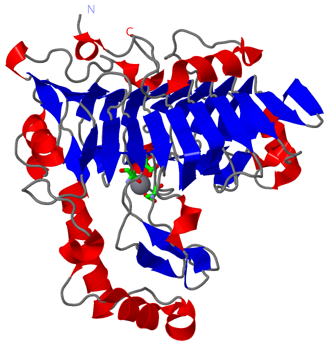
Asymmetric/Biological Unit (1, 1)
No.NameCountTypeFull Name
1CA1Ligand/IonCALCIUM ION

(-) Sites  (1, 1)

Asymmetric Unit (1, 1)
No.NameEvidenceResiduesDescription
1AC1SOFTWAREASP A:190 , ASP A:229 , ASP A:233 , HOH A:482 , HOH A:770 , HOH A:771BINDING SITE FOR RESIDUE CA A 417

(-) SS Bonds  (0, 0)

(no "SS Bond" information available for 1VBL)

(-) Cis Peptide Bonds  (1, 1)

Asymmetric/Biological Unit
No.Residues
1Leu A:286 -Pro A:287

 Sequence-Structure Mapping

(-) SAPs(SNPs)/Variants  (0, 0)

(no "SAP(SNP)/Variant" information available for 1VBL)

(-) PROSITE Motifs  (0, 0)

(no "PROSITE Motif" information available for 1VBL)

(-) Exons   (0, 0)

(no "Exon" information available for 1VBL)

(-) Sequences/Alignments

Asymmetric/Biological Unit
   Reformat: Number of residues per line =  ('0' or empty: single-line sequence representation)
  Number of residues per labelling interval =   
  UniProt sequence: complete  aligned part    
   Show mapping: SCOP domains CATH domains Pfam domains Secondary structure (by author)
SAPs(SNPs) PROSITE motifs Exons
(details for a mapped element are shown in a popup box when the mouse pointer rests over it)
Chain A from PDB  Type:PROTEIN  Length:416
 aligned with Q9AJM4_9BACI | Q9AJM4 from UniProtKB/TrEMBL  Length:441

    Alignment length:416
                                    35        45        55        65        75        85        95       105       115       125       135       145       155       165       175       185       195       205       215       225       235       245       255       265       275       285       295       305       315       325       335       345       355       365       375       385       395       405       415       425       435      
         Q9AJM4_9BACI    26 KELGHEVLKPYDGWAAYGEGTTGGAMASPQNVFVVTNRTELIQALGGNNHTNQYNSVPKIIYVKGTIDLNVDDNNQPVGPDFYKDPHFDFEAYLREYDPATWGKKEVEGPLEEARVRSQKKQKDRIMVYVGSNTSIIGVGKDAKIKGGGFLIKNVDNVIIRNIEFEAPLDYFPEWDPTDGTLGEWNSEYDSISIEGSSHIWIDHNTFTDGDHPDRSLGTYFGRPFQQHDGALDIKNSSDFITISYNVFTNHDKVTLIGASDSRMADSGHLRVTLHHNYYKNVTQRLPRVRFGQVHIYNNYYEFSNLADYDFQYAWGVGVFSQIYAQNNYFSFDWDIDPSLIIKVWSKNEESMYETGTIVDLPNGRRYIDLVASYNESNTLQLKKEVTWKPMFYHVIHPTPSVPALVKAKAGAGNLH 441
               SCOP domains d1vbla_ A: automated matches                                                                                                                                                                                                                                                                                                                                                                                                     SCOP domains
               CATH domains 1vblA00 A:1-416 Single-stranded right-handed beta-helix, Pectin lyase-like                                                                                                                                                                                                                                                                                                                                                       CATH domains
               Pfam domains --------------------------------------------------------------------------------------------------------------Pec_lyase_C-1vblA01 A:111-330                                                                                                                                                                                               -------------------------------------------------------------------------------------- Pfam domains
         Sec.struct. author .hhhhh......hhhhh..........hhh.eeee.hhhhhhhhh....hhhhhh...eeeee..eee..........hhhhhh....hhhhhhhhhhhhhhh.....hhhhhhhhhhhhhhhhhheee....eeeee.....eee..eeeee..eeeeee..eee.......eee.......eee.....eeee..eeeeee..eee....hhhhh.ee..ee.......eeee...eeeeee..eeeeeee..ee.....hhhhh....eeee..eeeeeee...ee...eeeee..eeee...........eeee...eeeee..eeee....hhhh.eeee.....eeeee..eeee..eeee.hhhhhhhhhh....ee.............hhhhhhhhhhhhh...... Sec.struct. author
                 SAPs(SNPs) -------------------------------------------------------------------------------------------------------------------------------------------------------------------------------------------------------------------------------------------------------------------------------------------------------------------------------------------------------------------------------------------------------------------------------- SAPs(SNPs)
                    PROSITE -------------------------------------------------------------------------------------------------------------------------------------------------------------------------------------------------------------------------------------------------------------------------------------------------------------------------------------------------------------------------------------------------------------------------------- PROSITE
                 Transcript -------------------------------------------------------------------------------------------------------------------------------------------------------------------------------------------------------------------------------------------------------------------------------------------------------------------------------------------------------------------------------------------------------------------------------- Transcript
                 1vbl A   1 KELGHEVLKPYDGWAAYGEGTTGGAMASPQNVFVVTNRTELIQALGGNNHTNQYNSVPKIIYVKGTIDLNVDDNNQPVGPDFYKDPHFDFEAYLREYDPATWGKKEVEGPLEEARVRSQKKQKDRIMVYVGSNTSIIGVGKDAKIKGGGFLIKNVDNVIIRNIEFEAPLDYFPEWDPTDGTLGEWNSEYDSISIEGSSHIWIDHNTFTDGDHPDRSLGTYFGRPFQQHDGALDIKNSSDFITISYNVFTNHDKVTLIGASDSRMADSGHLRVTLHHNYYKNVTQRLPRVRFGQVHIYNNYYEFSNLADYDFQYAWGVGVFSQIYAQNNYFSFDWDIDPSLIIKVWSKNEESMYETGTIVDLPNGRRYIDLVASYNESNTLQLKKEVTWKPMFYHVIHPTPSVPALVKAKAGAGNLH 416
                                    10        20        30        40        50        60        70        80        90       100       110       120       130       140       150       160       170       180       190       200       210       220       230       240       250       260       270       280       290       300       310       320       330       340       350       360       370       380       390       400       410      

   Legend:   → Mismatch (orange background)
  - → Gap (green background, '-', border residues have a numbering label)
    → Modified Residue (blue background, lower-case, 'x' indicates undefined single-letter code, labelled with number + name)
  x → Chemical Group (purple background, 'x', labelled with number + name, e.g. ACE or NH2)
  extra numbering lines below/above indicate numbering irregularities and modified residue names etc., number ends below/above '|'

 Classification and Annotation

(-) SCOP Domains  (1, 1)

Asymmetric/Biological Unit

(-) CATH Domains  (1, 1)

Asymmetric/Biological Unit
(-)
Class: Mainly Beta (13760)

(-) Pfam Domains  (1, 1)

Asymmetric/Biological Unit

(-) Gene Ontology  (5, 5)

Asymmetric/Biological Unit(hide GO term definitions)
Chain A   (Q9AJM4_9BACI | Q9AJM4)
molecular function
    GO:0016829    lyase activity    Catalysis of the cleavage of C-C, C-O, C-N and other bonds by other means than by hydrolysis or oxidation, or conversely adding a group to a double bond. They differ from other enzymes in that two substrates are involved in one reaction direction, but only one in the other direction. When acting on the single substrate, a molecule is eliminated and this generates either a new double bond or a new ring.
    GO:0046872    metal ion binding    Interacting selectively and non-covalently with any metal ion.
biological process
    GO:0005975    carbohydrate metabolic process    The chemical reactions and pathways involving carbohydrates, any of a group of organic compounds based of the general formula Cx(H2O)y. Includes the formation of carbohydrate derivatives by the addition of a carbohydrate residue to another molecule.
    GO:0000272    polysaccharide catabolic process    The chemical reactions and pathways resulting in the breakdown of a polysaccharide, a polymer of many (typically more than 10) monosaccharide residues linked glycosidically.
cellular component
    GO:0005576    extracellular region    The space external to the outermost structure of a cell. For cells without external protective or external encapsulating structures this refers to space outside of the plasma membrane. This term covers the host cell environment outside an intracellular parasite.

 Visualization

(-) Interactive Views

Asymmetric/Biological Unit
  Complete Structure
    Jena3D(integrated viewing of ligand, site, SAP, PROSITE, SCOP information)
    WebMol | AstexViewer[tm]@PDBe
(Java Applets, require no local installation except for Java; loading may be slow)
    STRAP
(Java WebStart application, automatic local installation, requires Java; full application with system access!)
    RasMol
(require local installation)
    Molscript (VRML)
(requires installation of a VRML viewer; select preferred view via VRML and generate a mono or stereo PDF format file)
 
  Ligands, Modified Residues, Ions
    CA  [ RasMol | Jena3D ]  +environment [ RasMol | Jena3D ]
 
  Sites
    AC1  [ RasMol ]  +environment [ RasMol ]
 
  Cis Peptide Bonds
    Leu A:286 - Pro A:287   [ RasMol ]  
 

(-) Still Images

Jmol
  protein: cartoon or spacefill or dots and stick; nucleic acid: cartoon and stick; ligands: spacefill; active site: stick
Molscript
  protein, nucleic acid: cartoon; ligands: spacefill; active site: ball and stick

 Databases and Analysis Tools

(-) Databases

Access by PDB/NDB ID
  1vbl
    Family and Domain InformationProDom | SYSTERS
    General Structural InformationGlycoscienceDB | MMDB | NDB | OCA | PDB | PDBe | PDBj | PDBsum | PDBWiki | PQS | PROTEOPEDIA
    Orientation in MembranesOPM
    Protein SurfaceSURFACE
    Secondary StructureDSSP (structure derived) | HSSP (homology derived)
    Structural GenomicsGeneCensus
    Structural NeighboursCE | VAST
    Structure ClassificationCATH | Dali | SCOP
    Validation and Original DataBMRB Data View | BMRB Restraints Grid | EDS | PROCHECK | RECOORD | WHAT_CHECK
 
Access by UniProt ID/Accession number
  Q9AJM4_9BACI | Q9AJM4
    Comparative Protein Structure ModelsModBase
    Genomic InformationEnsembl
    Protein-protein InteractionDIP
    Sequence, Family and Domain InformationInterPro | Pfam | SMART | UniProtKB/TrEMBL
 
Access by Enzyme Classificator   (EC Number)
  4.2.2.2
    General Enzyme InformationBRENDA | EC-PDB | Enzyme | IntEnz
    PathwayKEGG | MetaCyc
 
Access by Disease Identifier   (MIM ID)
  (no 'MIM ID' available)
    Disease InformationOMIM
 
Access by GenAge ID
  (no 'GenAge ID' available)
    Age Related InformationGenAge

(-) Analysis Tools

Access by PDB/NDB ID
    Domain InformationXDom
    Interatomic Contacts of Structural UnitsCSU
    Ligand-protein ContactsLPC
    Protein CavitiescastP
    Sequence and Secondary StructurePDBCartoon
    Structure AlignmentSTRAP(Java WebStart application, automatic local installation, requires Java; full application with system access!)
    Structure and Sequence BrowserSTING
 
Access by UniProt ID/Accession number
  Q9AJM4_9BACI | Q9AJM4
    Protein Disorder PredictionDisEMBL | FoldIndex | GLOBPLOT (for more information see DisProt)

 Related Entries

(-) Entries Sharing at Least One Protein Chain (UniProt ID)

(no "Entries Sharing at Least One Protein Chain" available for 1VBL)

(-) Related Entries Specified in the PDB File

(no "Related Entries Specified in the PDB File" available for 1VBL)