Show PDB file:   
         Plain Text   HTML   (compressed file size)
QuickSearch:   
by PDB,NDB,UniProt,PROSITE Code or Search Term(s)  
(-)Asymmetric Unit
(-)Biological Unit 1
(-)Biological Unit 2
collapse expand < >
Image Asymmetric Unit
Asymmetric Unit  (Jmol Viewer)
Image Biological Unit 1
Biological Unit 1  (Jmol Viewer)
Image Biological Unit 2
Biological Unit 2  (Jmol Viewer)

(-) Description

Title :  THE MONOMERIC ASP49 SECRETORY PHOSPHOLIPASE A2 FROM THE VENOM OF AGKISTRIDON PISCIVORUS PISCIVORUS
 
Authors :  D. L. Scott
Date :  15 Nov 96  (Deposition) - 07 Jul 97  (Release) - 24 Feb 09  (Revision)
Method :  X-RAY DIFFRACTION
Resolution :  1.60
Chains :  Asym. Unit :  A,B
Biol. Unit 1:  A  (1x)
Biol. Unit 2:  B  (1x)
Keywords :  Phospholipase A2, Lipid Degradation, Hydrolase (Keyword Search: [Gene Ontology, PubMed, Web (Google))
 
Reference :  S. K. Han, E. T. Yoon, D. L. Scott, P. B. Sigler, W. Cho
Structural Aspects Of Interfacial Adsorption. A Crystallographic And Site-Directed Mutagenesis Study Of The Phospholipase A2 From The Venom Of Agkistrodon Piscivorus Piscivorus.
J. Biol. Chem. V. 272 3573 1997
PubMed-ID: 9013608  |  Reference-DOI: 10.1074/JBC.272.26.16152
(for further references see the PDB file header)

(-) Compounds

Molecule 1 - PHOSPHOLIPASE A2
    ChainsA, B
    EC Number3.1.1.4
    Organism CommonEASTERN COTTONMOUTH
    Organism ScientificAGKISTRODON PISCIVORUS PISCIVORUS
    Organism Taxid8716
    Other DetailsSECRETORY PHOSPHOLIPASE A2
    StrainPISCIVORUS

 Structural Features

(-) Chains, Units

  12
Asymmetric Unit AB
Biological Unit 1 (1x)A 
Biological Unit 2 (1x) B

Summary Information (see also Sequences/Alignments below)

(-) Ligands, Modified Residues, Ions  (0, 0)

(no "Ligand,Modified Residues,Ions" information available for 1VAP)

(-) Sites  (0, 0)

(no "Site" information available for 1VAP)
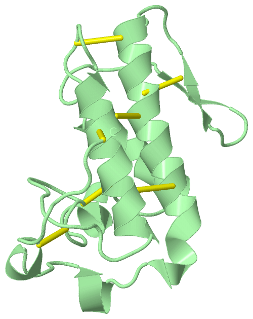
(-) SS Bonds  (14, 14)

Asymmetric Unit
No.Residues
1A:26 -A:116
2A:28 -A:44
3A:43 -A:95
4A:49 -A:123
5A:50 -A:88
6A:57 -A:81
7A:75 -A:86
8B:26 -B:116
9B:28 -B:44
10B:43 -B:95
11B:49 -B:123
12B:50 -B:88
13B:57 -B:81
14B:75 -B:86

(-) Cis Peptide Bonds  (0, 0)

(no "Cis Peptide Bond" information available for 1VAP)

 Sequence-Structure Mapping

(-) SAPs(SNPs)/Variants  (0, 0)

(no "SAP(SNP)/Variant" information available for 1VAP)

(-) PROSITE Motifs  (2, 4)

Asymmetric Unit (2, 4)
 PROSITEUniProtKBPDB
No.IDACDescriptionIDLocationCountLocation
1PA2_HISPS00118 Phospholipase A2 histidine active site.PA2B1_AGKPI43-50
 
  2A:43-50
B:43-50
2PA2_ASPPS00119 Phospholipase A2 aspartic acid active site.PA2B1_AGKPI85-95
 
  2A:85-95
B:85-95
Biological Unit 1 (2, 2)
 PROSITEUniProtKBPDB
No.IDACDescriptionIDLocationCountLocation
1PA2_HISPS00118 Phospholipase A2 histidine active site.PA2B1_AGKPI43-50
 
  1A:43-50
-
2PA2_ASPPS00119 Phospholipase A2 aspartic acid active site.PA2B1_AGKPI85-95
 
  1A:85-95
-
Biological Unit 2 (2, 2)
 PROSITEUniProtKBPDB
No.IDACDescriptionIDLocationCountLocation
1PA2_HISPS00118 Phospholipase A2 histidine active site.PA2B1_AGKPI43-50
 
  1-
B:43-50
2PA2_ASPPS00119 Phospholipase A2 aspartic acid active site.PA2B1_AGKPI85-95
 
  1-
B:85-95

(-) Exons   (0, 0)

(no "Exon" information available for 1VAP)

(-) Sequences/Alignments

Asymmetric Unit
   Reformat: Number of residues per line =  ('0' or empty: single-line sequence representation)
  Number of residues per labelling interval =   
  UniProt sequence: complete  aligned part    
   Show mapping: SCOP domains CATH domains Pfam domains Secondary structure (by author)
SAPs(SNPs) PROSITE motifs Exons
(details for a mapped element are shown in a popup box when the mouse pointer rests over it)
Chain A from PDB  Type:PROTEIN  Length:123
 aligned with PA2B1_AGKPI | P51972 from UniProtKB/Swiss-Prot  Length:123

    Alignment length:123
                                    10        20        30        40        50        60        70        80        90       100       110       120   
          PA2B1_AGKPI     1 NLFQFEKLIKKMTGKSGMLWYSAYGCYCGWGGQGRPKDATDRCCFVHDCCYGKVTGCNPKMDIYTYSVDNGNIVCGGTNPCKKQICECDRAAAICFRDNLKTYDSKTYWKYPKKNCKEESEPC 123
               SCOP domains d1vapa_ A: Snake phospholipase A2                                                                                           SCOP domains
               CATH domains 1vapA00 A:1-123 Phospholipase A2                                                                                            CATH domains
               Pfam domains --------------------------------------------------------------------------------------------------------------------------- Pfam domains
         Sec.struct. author .hhhhhhhhhhhh...hhhh..................hhhhhhhhhhhhhhh............eeee..eeee....hhhhhhhhhhhhhhhhhhh.hhh..hhhh....hhh........ Sec.struct. author
                 SAPs(SNPs) --------------------------------------------------------------------------------------------------------------------------- SAPs(SNPs)
                    PROSITE ------------------------------------------PA2_HIS ----------------------------------PA2_ASP    ---------------------------- PROSITE
                 Transcript --------------------------------------------------------------------------------------------------------------------------- Transcript
                 1vap A   1 NLFQFEKLIKKMTGKSGMLWYSAYGCYCGWGGQGRPKDATDRCCFVHDCCYGKVTGCNPKMDIYTYSVDNGNIVCGGTNPCKKQICECDRAAAICFRDNLKTYDSKTYWKYPKKNCKEESEPC 123
                                    10        20        30        40        50        60        70        80        90       100       110       120   

Chain B from PDB  Type:PROTEIN  Length:123
 aligned with PA2B1_AGKPI | P51972 from UniProtKB/Swiss-Prot  Length:123

    Alignment length:123
                                    10        20        30        40        50        60        70        80        90       100       110       120   
          PA2B1_AGKPI     1 NLFQFEKLIKKMTGKSGMLWYSAYGCYCGWGGQGRPKDATDRCCFVHDCCYGKVTGCNPKMDIYTYSVDNGNIVCGGTNPCKKQICECDRAAAICFRDNLKTYDSKTYWKYPKKNCKEESEPC 123
               SCOP domains d1vapb_ B: Snake phospholipase A2                                                                                           SCOP domains
               CATH domains 1vapB00 B:1-123 Phospholipase A2                                                                                            CATH domains
               Pfam domains --------------------------------------------------------------------------------------------------------------------------- Pfam domains
         Sec.struct. author .hhhhhhhhhhhh...hhhhh.................hhhhhhhhhhhhhhh............eee....eee....hhhhhhhhhhhhhhhhhhhhhhh..hhhh....hhh........ Sec.struct. author
                 SAPs(SNPs) --------------------------------------------------------------------------------------------------------------------------- SAPs(SNPs)
                    PROSITE ------------------------------------------PA2_HIS ----------------------------------PA2_ASP    ---------------------------- PROSITE
                 Transcript --------------------------------------------------------------------------------------------------------------------------- Transcript
                 1vap B   1 NLFQFEKLIKKMTGKSGMLWYSAYGCYCGWGGQGRPKDATDRCCFVHDCCYGKVTGCNPKMDIYTYSVDNGNIVCGGTNPCKKQICECDRAAAICFRDNLKTYDSKTYWKYPKKNCKEESEPC 123
                                    10        20        30        40        50        60        70        80        90       100       110       120   

   Legend:   → Mismatch (orange background)
  - → Gap (green background, '-', border residues have a numbering label)
    → Modified Residue (blue background, lower-case, 'x' indicates undefined single-letter code, labelled with number + name)
  x → Chemical Group (purple background, 'x', labelled with number + name, e.g. ACE or NH2)
  extra numbering lines below/above indicate numbering irregularities and modified residue names etc., number ends below/above '|'

 Classification and Annotation

(-) SCOP Domains  (1, 2)

Asymmetric Unit

(-) CATH Domains  (1, 2)

Asymmetric Unit

(-) Pfam Domains  (0, 0)

(no "Pfam Domain" information available for 1VAP)

(-) Gene Ontology  (7, 7)

Asymmetric Unit(hide GO term definitions)
Chain A,B   (PA2B1_AGKPI | P51972)
molecular function
    GO:0005509    calcium ion binding    Interacting selectively and non-covalently with calcium ions (Ca2+).
    GO:0016787    hydrolase activity    Catalysis of the hydrolysis of various bonds, e.g. C-O, C-N, C-C, phosphoric anhydride bonds, etc. Hydrolase is the systematic name for any enzyme of EC class 3.
    GO:0046872    metal ion binding    Interacting selectively and non-covalently with any metal ion.
    GO:0004623    phospholipase A2 activity    Catalysis of the reaction: phosphatidylcholine + H2O = 1-acylglycerophosphocholine + a carboxylate.
biological process
    GO:0016042    lipid catabolic process    The chemical reactions and pathways resulting in the breakdown of lipids, compounds soluble in an organic solvent but not, or sparingly, in an aqueous solvent.
    GO:0006629    lipid metabolic process    The chemical reactions and pathways involving lipids, compounds soluble in an organic solvent but not, or sparingly, in an aqueous solvent. Includes fatty acids; neutral fats, other fatty-acid esters, and soaps; long-chain (fatty) alcohols and waxes; sphingoids and other long-chain bases; glycolipids, phospholipids and sphingolipids; and carotenes, polyprenols, sterols, terpenes and other isoprenoids.
cellular component
    GO:0005576    extracellular region    The space external to the outermost structure of a cell. For cells without external protective or external encapsulating structures this refers to space outside of the plasma membrane. This term covers the host cell environment outside an intracellular parasite.

 Visualization

(-) Interactive Views

Asymmetric Unit
  Complete Structure
    Jena3D(integrated viewing of ligand, site, SAP, PROSITE, SCOP information)
    WebMol | AstexViewer[tm]@PDBe
(Java Applets, require no local installation except for Java; loading may be slow)
    STRAP
(Java WebStart application, automatic local installation, requires Java; full application with system access!)
    RasMol
(require local installation)
    Molscript (VRML)
(requires installation of a VRML viewer; select preferred view via VRML and generate a mono or stereo PDF format file)
 
  Ligands, Modified Residues, Ions
(no "Ligands, Modified Residues, Ions" information available for 1vap)
 
  Sites
(no "Sites" information available for 1vap)
 
  Cis Peptide Bonds
(no "Cis Peptide Bonds" information available for 1vap)
 
Biological Units
  Complete Structure
    Biological Unit 1  [ Jena3D ]
    Biological Unit 2  [ Jena3D ]

(-) Still Images

Jmol
  protein: cartoon or spacefill or dots and stick; nucleic acid: cartoon and stick; ligands: spacefill; active site: stick
Molscript
  protein, nucleic acid: cartoon; ligands: spacefill; active site: ball and stick

 Databases and Analysis Tools

(-) Databases

Access by PDB/NDB ID
  1vap
    Family and Domain InformationProDom | SYSTERS
    General Structural InformationGlycoscienceDB | MMDB | NDB | OCA | PDB | PDBe | PDBj | PDBsum | PDBWiki | PQS | PROTEOPEDIA
    Orientation in MembranesOPM
    Protein SurfaceSURFACE
    Secondary StructureDSSP (structure derived) | HSSP (homology derived)
    Structural GenomicsGeneCensus
    Structural NeighboursCE | VAST
    Structure ClassificationCATH | Dali | SCOP
    Validation and Original DataBMRB Data View | BMRB Restraints Grid | EDS | PROCHECK | RECOORD | WHAT_CHECK
 
Access by UniProt ID/Accession number
  PA2B1_AGKPI | P51972
    Comparative Protein Structure ModelsModBase
    Genomic InformationEnsembl
    Protein-protein InteractionDIP
    Sequence, Family and Domain InformationInterPro | Pfam | SMART | UniProtKB/SwissProt
 
Access by Enzyme Classificator   (EC Number)
  3.1.1.4
    General Enzyme InformationBRENDA | EC-PDB | Enzyme | IntEnz
    PathwayKEGG | MetaCyc
 
Access by Disease Identifier   (MIM ID)
  (no 'MIM ID' available)
    Disease InformationOMIM
 
Access by GenAge ID
  (no 'GenAge ID' available)
    Age Related InformationGenAge

(-) Analysis Tools

Access by PDB/NDB ID
    Domain InformationXDom
    Interatomic Contacts of Structural UnitsCSU
    Ligand-protein ContactsLPC
    Protein CavitiescastP
    Sequence and Secondary StructurePDBCartoon
    Structure AlignmentSTRAP(Java WebStart application, automatic local installation, requires Java; full application with system access!)
    Structure and Sequence BrowserSTING
 
Access by UniProt ID/Accession number
  PA2B1_AGKPI | P51972
    Protein Disorder PredictionDisEMBL | FoldIndex | GLOBPLOT (for more information see DisProt)

 Related Entries

(-) Entries Sharing at Least One Protein Chain (UniProt ID)

(no "Entries Sharing at Least One Protein Chain" available for 1VAP)

(-) Related Entries Specified in the PDB File

(no "Related Entries Specified in the PDB File" available for 1VAP)