Show PDB file:   
         Plain Text   HTML   (compressed file size)
QuickSearch:   
by PDB,NDB,UniProt,PROSITE Code or Search Term(s)  
(-)NMR Structure - manually
(-)NMR Structure - model 1
(-)NMR Structure - model 1, sites
(-)NMR Structure - all models
collapse expand < >
Image NMR Structure - manually
NMR Structure - manually  (Jmol Viewer)
Image NMR Structure - model 1
NMR Structure - model 1  (Jmol Viewer)
Image NMR Structure - model 1, sites
NMR Structure - model 1, sites  (Jmol Viewer)
Image NMR Structure - all models
NMR Structure - all models  (Jmol Viewer)

(-) Description

Title :  SOLUTION STRUCTURE OF THE FIRST ZN-FINGER DOMAIN OF POLY(ADP-RIBOSE) POLYMERASE-1
 
Authors :  T. Nagashima, F. Hayashi, S. Yokoyama, Riken Structural Genomics/Proteomics Initiative (Rsgi)
Date :  04 Feb 04  (Deposition) - 22 Feb 05  (Release) - 24 Feb 09  (Revision)
Method :  SOLUTION NMR
Resolution :  NOT APPLICABLE
Chains :  NMR Structure  :  A  (20x)
Keywords :  Parp, Dna Repair, Inflammation, Cell Death, Structural Genomics, Riken Structural Genomics/Proteomics Initiative, Rsgi, Transferase (Keyword Search: [Gene Ontology, PubMed, Web (Google))
 
Reference :  T. Nagashima, F. Hayashi, S. Yokoyama
Solution Structure Of The First Zn-Finger Domain Of Poly(Adp-Ribose) Polymerase-1
To Be Published
PubMed: search
(for further references see the PDB file header)

(-) Compounds

Molecule 1 - POLY (ADP-RIBOSE) POLYMERASE
    ChainsA
    EC Number2.4.2.30
    EngineeredYES
    Expression System PlasmidP021118-12
    Expression System Vector TypePLASMID
    FragmentZN-FINGER DOMAIN
    GeneRIKEN CDNA RAFL09-59-L22
    Organism CommonTHALE CRESS
    Organism ScientificARABIDOPSIS THALIANA
    Organism Taxid3702
    Other DetailsCELL-FREE PROTEIN SYNTHESIS

 Structural Features

(-) Chains, Units

  
NMR Structure (20x)

Summary Information (see also Sequences/Alignments below)

(-) Ligands, Modified Residues, Ions  (1, 1)

NMR Structure (1, 1)
No.NameCountTypeFull Name
1ZN1Ligand/IonZINC ION

(-) Sites  (1, 1)

NMR Structure (1, 1)
No.NameEvidenceResiduesDescription
1AC1SOFTWARECYS A:23 , CYS A:26 , HIS A:55 , CYS A:58BINDING SITE FOR RESIDUE ZN A 200

(-) SS Bonds  (0, 0)

(no "SS Bond" information available for 1V9X)

(-) Cis Peptide Bonds  (0, 0)

(no "Cis Peptide Bond" information available for 1V9X)

 Sequence-Structure Mapping

(-) SAPs(SNPs)/Variants  (0, 0)

(no "SAP(SNP)/Variant" information available for 1V9X)

(-) PROSITE Motifs  (1, 2)

NMR Structure (1, 2)
 PROSITEUniProtKBPDB
No.IDACDescriptionIDLocationCountLocation
1PARP_ZN_FINGER_2PS50064 Poly(ADP-ribose) polymerase zinc finger domain profile.PARP1_ARATH8-91
114-186
  2A:11-94
A:110-114

(-) Exons   (0, 0)

(no "Exon" information available for 1V9X)

(-) Sequences/Alignments

NMR Structure
   Reformat: Number of residues per line =  ('0' or empty: single-line sequence representation)
  Number of residues per labelling interval =   
  UniProt sequence: complete  aligned part    
   Show mapping: SCOP domains CATH domains Pfam domains Secondary structure (by author)
SAPs(SNPs) PROSITE motifs Exons
(details for a mapped element are shown in a popup box when the mouse pointer rests over it)
Chain A from PDB  Type:PROTEIN  Length:114
 aligned with PARP1_ARATH | Q9ZP54 from UniProtKB/Swiss-Prot  Length:983

    Alignment length:177
                               1                                                                                                                                                                             
                               |     7        17        27        37        47        57        67        77        87        97       107       117       127       137       147       157       167       
          PARP1_ARATH     - ---MASPHKPWRAEYAKSSRSSCKTCKSVINKENFRLGKLVQSTHFDGIMPMWNHASCILKKTKQIKSVDDVEGIESLRWEDQQKIRKYVESGAGSNTSTSTGTSTSSTANNAKLEYGIEVSQTSRAGCRKCSEKILKGEVRIFSKPEGPGNKGLMWHHAKCFLEMSSSTELESLSG 174
               SCOP domains --------------------------------------------------------------------------------------------------------------------------------------------------------------------------------- SCOP domains
               CATH domains 1v9xA00 A:1-114 first zn-finger domain of poly(adp-ribose) polymerase-1                                                                                                           CATH domains
               Pfam domains -------------zf-PARP-1v9xA01 A:14-91                                                       -------------------------------------------------------------------------------------- Pfam domains
         Sec.struct. author ..........eeeee...................eeeee............eeeehhhhhh......hhh.ee.....hhhhhhhhhhhhh..................---------------------------------------...------------------------.. Sec.struct. author
                 SAPs(SNPs) --------------------------------------------------------------------------------------------------------------------------------------------------------------------------------- SAPs(SNPs)
                    PROSITE ----------PARP_ZN_FINGER_2  PDB: A:11-94 UniProt: 8-91                                        ----------------------PARP_ZN_FINGER_2  PDB: A:110-114 UniProt: 114-186             PROSITE
                 Transcript --------------------------------------------------------------------------------------------------------------------------------------------------------------------------------- Transcript
                 1v9x A   1 GSSGSSGHKPWRAEYAKSSRSSCKTCKSVINKENFRLGKLVQSTHFDGIMPMWNHASCILKKTKQIKSVDDVEGIESLRWEDQQKIRKYVESGAGSNTSTSTGTSTSSS---------------------------------------GPS------------------------SG 114
                                    10        20        30        40        50        60        70        80        90       100        |-         -         -         -       111|        -         -     | 
                                                                                                                                      109                                     110 |                      113 
                                                                                                                                                                                112                          

   Legend:   → Mismatch (orange background)
  - → Gap (green background, '-', border residues have a numbering label)
    → Modified Residue (blue background, lower-case, 'x' indicates undefined single-letter code, labelled with number + name)
  x → Chemical Group (purple background, 'x', labelled with number + name, e.g. ACE or NH2)
  extra numbering lines below/above indicate numbering irregularities and modified residue names etc., number ends below/above '|'

 Classification and Annotation

(-) SCOP Domains  (0, 0)

(no "SCOP Domain" information available for 1V9X)

(-) CATH Domains  (1, 1)

NMR Structure
(-)
Class: Alpha Beta (26913)

(-) Pfam Domains  (1, 1)

NMR Structure

(-) Gene Ontology  (16, 16)

NMR Structure(hide GO term definitions)
Chain A   (PARP1_ARATH | Q9ZP54)
molecular function
    GO:0003677    DNA binding    Any molecular function by which a gene product interacts selectively and non-covalently with DNA (deoxyribonucleic acid).
    GO:0003910    DNA ligase (ATP) activity    Catalysis of the reaction: ATP + deoxyribonucleotide(n) + deoxyribonucleotide(m) = AMP + diphosphate + deoxyribonucleotide(n+m).
    GO:0051287    NAD binding    Interacting selectively and non-covalently with nicotinamide adenine dinucleotide, a coenzyme involved in many redox and biosynthetic reactions; binding may be to either the oxidized form, NAD+, or the reduced form, NADH.
    GO:0003950    NAD+ ADP-ribosyltransferase activity    Catalysis of the reaction: NAD+ + (ADP-D-ribosyl)(n)-acceptor = nicotinamide + (ADP-D-ribosyl)(n+1)-acceptor.
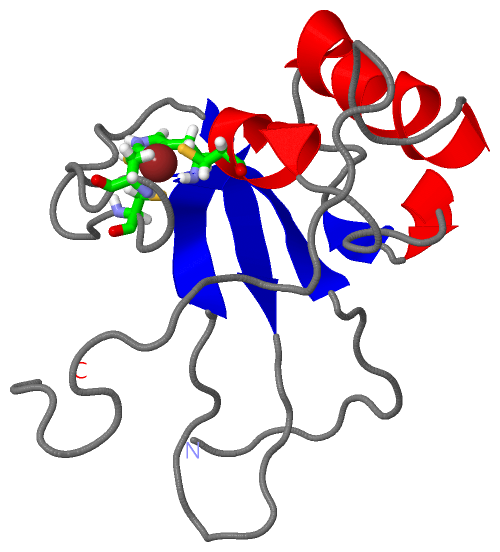
    GO:0046872    metal ion binding    Interacting selectively and non-covalently with any metal ion.
    GO:0016740    transferase activity    Catalysis of the transfer of a group, e.g. a methyl group, glycosyl group, acyl group, phosphorus-containing, or other groups, from one compound (generally regarded as the donor) to another compound (generally regarded as the acceptor). Transferase is the systematic name for any enzyme of EC class 2.
    GO:0016757    transferase activity, transferring glycosyl groups    Catalysis of the transfer of a glycosyl group from one compound (donor) to another (acceptor).
    GO:0008270    zinc ion binding    Interacting selectively and non-covalently with zinc (Zn) ions.
biological process
    GO:0051103    DNA ligation involved in DNA repair    The re-formation of a broken phosphodiester bond in the DNA backbone, carried out by DNA ligase, that contributes to DNA repair.
    GO:0006281    DNA repair    The process of restoring DNA after damage. Genomes are subject to damage by chemical and physical agents in the environment (e.g. UV and ionizing radiations, chemical mutagens, fungal and bacterial toxins, etc.) and by free radicals or alkylating agents endogenously generated in metabolism. DNA is also damaged because of errors during its replication. A variety of different DNA repair pathways have been reported that include direct reversal, base excision repair, nucleotide excision repair, photoreactivation, bypass, double-strand break repair pathway, and mismatch repair pathway.
    GO:0006273    lagging strand elongation    The synthesis of DNA from a template strand in a net 3' to 5' direction. Lagging strand DNA elongation proceeds by discontinuous synthesis of short stretches of DNA, known as Okazaki fragments, from RNA primers; these fragments are then joined by DNA ligase. Although each segment of nascent DNA is synthesized in the 5' to 3' direction, the overall direction of lagging strand synthesis is 3' to 5', mirroring the progress of the replication fork.
    GO:0006471    protein ADP-ribosylation    The transfer, from NAD, of ADP-ribose to protein amino acids.
    GO:0009737    response to abscisic acid    Any process that results in a change in state or activity of a cell or an organism (in terms of movement, secretion, enzyme production, gene expression, etc.) as a result of an abscisic acid stimulus.
    GO:0006979    response to oxidative stress    Any process that results in a change in state or activity of a cell or an organism (in terms of movement, secretion, enzyme production, gene expression, etc.) as a result of oxidative stress, a state often resulting from exposure to high levels of reactive oxygen species, e.g. superoxide anions, hydrogen peroxide (H2O2), and hydroxyl radicals.
cellular component
    GO:0005737    cytoplasm    All of the contents of a cell excluding the plasma membrane and nucleus, but including other subcellular structures.
    GO:0005634    nucleus    A membrane-bounded organelle of eukaryotic cells in which chromosomes are housed and replicated. In most cells, the nucleus contains all of the cell's chromosomes except the organellar chromosomes, and is the site of RNA synthesis and processing. In some species, or in specialized cell types, RNA metabolism or DNA replication may be absent.
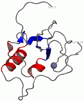
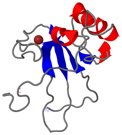
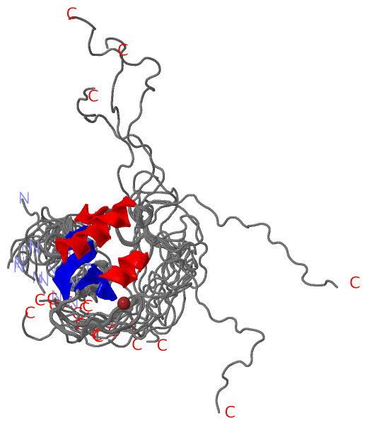
 Visualization

(-) Interactive Views

NMR Structure
  Complete Structure
    Jena3D(integrated viewing of ligand, site, SAP, PROSITE, SCOP information)
    WebMol | AstexViewer[tm]@PDBe
(Java Applets, require no local installation except for Java; loading may be slow)
    STRAP
(Java WebStart application, automatic local installation, requires Java; full application with system access!)
    RasMol
(require local installation)
    Molscript (VRML)
(requires installation of a VRML viewer; select preferred view via VRML and generate a mono or stereo PDF format file)
 
  Ligands, Modified Residues, Ions
    ZN  [ RasMol | Jena3D ]  +environment [ RasMol | Jena3D ]
 
  Sites
    AC1  [ RasMol ]  +environment [ RasMol ]
 
  Cis Peptide Bonds
(no "Cis Peptide Bonds" information available for 1v9x)
 

(-) Still Images

Jmol
  protein: cartoon or spacefill or dots and stick; nucleic acid: cartoon and stick; ligands: spacefill; active site: stick
Molscript
  protein, nucleic acid: cartoon; ligands: spacefill; active site: ball and stick

 Databases and Analysis Tools

(-) Databases

Access by PDB/NDB ID
  1v9x
    Family and Domain InformationProDom | SYSTERS
    General Structural InformationGlycoscienceDB | MMDB | NDB | OCA | PDB | PDBe | PDBj | PDBsum | PDBWiki | PQS | PROTEOPEDIA
    Orientation in MembranesOPM
    Protein SurfaceSURFACE
    Secondary StructureDSSP (structure derived) | HSSP (homology derived)
    Structural GenomicsGeneCensus
    Structural NeighboursCE | VAST
    Structure ClassificationCATH | Dali | SCOP
    Validation and Original DataBMRB Data View | BMRB Restraints Grid | EDS | PROCHECK | RECOORD | WHAT_CHECK
 
Access by UniProt ID/Accession number
  PARP1_ARATH | Q9ZP54
    Comparative Protein Structure ModelsModBase
    Genomic InformationEnsembl
    Protein-protein InteractionDIP
    Sequence, Family and Domain InformationInterPro | Pfam | SMART | UniProtKB/SwissProt
 
Access by Enzyme Classificator   (EC Number)
  2.4.2.30
    General Enzyme InformationBRENDA | EC-PDB | Enzyme | IntEnz
    PathwayKEGG | MetaCyc
 
Access by Disease Identifier   (MIM ID)
  (no 'MIM ID' available)
    Disease InformationOMIM
 
Access by GenAge ID
  (no 'GenAge ID' available)
    Age Related InformationGenAge

(-) Analysis Tools

Access by PDB/NDB ID
    Domain InformationXDom
    Interatomic Contacts of Structural UnitsCSU
    Ligand-protein ContactsLPC
    Protein CavitiescastP
    Sequence and Secondary StructurePDBCartoon
    Structure AlignmentSTRAP(Java WebStart application, automatic local installation, requires Java; full application with system access!)
    Structure and Sequence BrowserSTING
 
Access by UniProt ID/Accession number
  PARP1_ARATH | Q9ZP54
    Protein Disorder PredictionDisEMBL | FoldIndex | GLOBPLOT (for more information see DisProt)

 Related Entries

(-) Entries Sharing at Least One Protein Chain (UniProt ID)

(no "Entries Sharing at Least One Protein Chain" available for 1V9X)

(-) Related Entries Specified in the PDB File

(no "Related Entries Specified in the PDB File" available for 1V9X)