Show PDB file:   
         Plain Text   HTML   (compressed file size)
QuickSearch:   
by PDB,NDB,UniProt,PROSITE Code or Search Term(s)  
(-)NMR Structure - model 1
(-)NMR Structure - all models
collapse expand < >
Image NMR Structure - model 1
NMR Structure - model 1  (Jmol Viewer)
Image NMR Structure - all models
NMR Structure - all models  (Jmol Viewer)

(-) Description

Title :  STERILE ALPHA MOTIF (SAM) DOMAIN OF MOUSE BIFUNCTIONAL APOPTOSIS REGULATOR
 
Authors :  A. Goroncy, T. Kigawa, S. Koshiba, F. Hayashi, N. Kobayashi, N. Tochio, M. Inoue, S. Yokoyama, Riken Structural Genomics/Proteomics Initiative (Rsgi)
Date :  29 Dec 03  (Deposition) - 25 Jan 05  (Release) - 24 Feb 09  (Revision)
Method :  SOLUTION NMR
Resolution :  NOT APPLICABLE
Chains :  NMR Structure  :  A  (20x)
Keywords :  Apoptosis, Neuron, Cell Death, Structural Genomics, Riken Structural Genomics/Proteomics Initiative, Rsgi (Keyword Search: [Gene Ontology, PubMed, Web (Google))
 
Reference :  A. Goroncy, T. Kigawa, S. Koshiba, F. Hayashi, N. Kobayashi, N. Tochio, M. Inoue, S. Yokoyama
Sterile Alpha Motif (Sam) Domain Of Mouse Bifunctional Apoptosis Regulator
To Be Published
PubMed: search
(for further references see the PDB file header)

(-) Compounds

Molecule 1 - SIMILAR TO RING FINGER PROTEIN 36
    ChainsA
    EngineeredYES
    Expression System PlasmidP030120-59
    Expression System Vector TypePLASMID
    FragmentSTERILE ALPHA MOTIF (SAM) DOMAIN
    GeneRIKEN CDNA 3010001A07
    Organism CommonHOUSE MOUSE
    Organism ScientificMUS MUSCULUS
    Organism Taxid10090
    Other DetailsCELL-FREE PROTEIN SYNTHESIS
    SynonymBIFUNCTIONAL APOPTOSIS REGULATOR

 Structural Features

(-) Chains, Units

  
NMR Structure (20x)

Summary Information (see also Sequences/Alignments below)

(-) Ligands, Modified Residues, Ions  (0, 0)

(no "Ligand,Modified Residues,Ions" information available for 1V85)

(-) Sites  (0, 0)

(no "Site" information available for 1V85)

(-) SS Bonds  (0, 0)

(no "SS Bond" information available for 1V85)

(-) Cis Peptide Bonds  (1, 20)

NMR Structure
No.ModelResidues
11, 2, 3, 4, 5, 6, 7, 8, 9, 10, 11, 12, 13, 14, 15, 16, 17, 18, 19, 20Ala A:64 -Pro A:65

 Sequence-Structure Mapping

(-) SAPs(SNPs)/Variants  (0, 0)

(no "SAP(SNP)/Variant" information available for 1V85)

(-) PROSITE Motifs  (1, 1)

NMR Structure (1, 1)
 PROSITEUniProtKBPDB
No.IDACDescriptionIDLocationCountLocation
1SAM_DOMAINPS50105 SAM domain profile.BFAR_MOUSE182-249  1A:20-85

(-) Exons   (0, 0)

(no "Exon" information available for 1V85)

(-) Sequences/Alignments

NMR Structure
   Reformat: Number of residues per line =  ('0' or empty: single-line sequence representation)
  Number of residues per labelling interval =   
  UniProt sequence: complete  aligned part    
   Show mapping: SCOP domains CATH domains Pfam domains Secondary structure (by author)
SAPs(SNPs) PROSITE motifs Exons
(details for a mapped element are shown in a popup box when the mouse pointer rests over it)
Chain A from PDB  Type:PROTEIN  Length:91
 aligned with BFAR_MOUSE | Q8R079 from UniProtKB/Swiss-Prot  Length:450

    Alignment length:189
                                   132       142       152       162       172       182       192       202       212       222       232       242       252       262       272       282       292       302         
           BFAR_MOUSE   123 GNDQNPLAPSTGRVNPQRGGGFFSGVLTALTGVAVILLVYHWRSRESEHGLLVHKAVDKWTMEEVVLWLEQLGPWASLYRDRFLSERVNGRLLLTLTEEEFSRAPYTIENSSHRRVILTELERVRALGVKPPQNLWEYKAVNPGRSLFLLYALKSSPRLGLLYLYLFDYTDCFLPFIHTICPLQENSSG 311
               SCOP domains --------------------------------------------------------------------------------------------------------------------------------------------------------------------------------------------- SCOP domains
               CATH domains --------------------------------------------------------------------------------------------------------------------------------------------------------------------------------------------- CATH domains
               Pfam domains ---------------------------------------------------------SAM_1-1v85A01 A:18-85                                               ---------------------------------------------------------------- Pfam domains
         Sec.struct. author .--------...-----------.------..---------------..hhhhhhhhhhhhhhhhhhhhhhhh..hhhhhhhhhhh..hhhhhhhhhhhhhhh......hhhhhhhhhhhhhhhh-----------------------------h..-----------------------------... Sec.struct. author
                 SAPs(SNPs) --------------------------------------------------------------------------------------------------------------------------------------------------------------------------------------------- SAPs(SNPs)
                    PROSITE -----------------------------------------------------------SAM_DOMAIN  PDB: A:20-85 UniProt: 182-249                           -------------------------------------------------------------- PROSITE
                 Transcript --------------------------------------------------------------------------------------------------------------------------------------------------------------------------------------------- Transcript
                 1v85 A   1 G--------SSG-----------S------SG---------------EHGLLVHKAVDKWTTEEVVLWLEQLGPWASLYRDRFLSERVNGRLLLTLTEEEFSRAPYTIENSSHRRVILTELERVR-----------------------------SGP-----------------------------SSG  91
                            |        2 |       -   |     -||       -       |10        20        30        40        50        60        70        80    |    -         -         -    | |  -         -         -      |  
                            |        2 4           5      6|               8                                                                           85                            86 |                            89  
                            1                              7                                                                                                                           88                                

   Legend:   → Mismatch (orange background)
  - → Gap (green background, '-', border residues have a numbering label)
    → Modified Residue (blue background, lower-case, 'x' indicates undefined single-letter code, labelled with number + name)
  x → Chemical Group (purple background, 'x', labelled with number + name, e.g. ACE or NH2)
  extra numbering lines below/above indicate numbering irregularities and modified residue names etc., number ends below/above '|'

 Classification and Annotation

(-) SCOP Domains  (0, 0)

(no "SCOP Domain" information available for 1V85)

(-) CATH Domains  (0, 0)

(no "CATH Domain" information available for 1V85)

(-) Pfam Domains  (1, 1)

NMR Structure
(-)
Clan: SAM (26)

(-) Gene Ontology  (18, 18)

NMR Structure(hide GO term definitions)
Chain A   (BFAR_MOUSE | Q8R079)
molecular function
    GO:0089720    caspase binding    Interacting selectively and non-covalently with a caspase family protein.
    GO:0046872    metal ion binding    Interacting selectively and non-covalently with any metal ion.
    GO:0030674    protein binding, bridging    The binding activity of a molecule that brings together two or more protein molecules, or a protein and another macromolecule or complex, through a selective, non-covalent, often stoichiometric interaction, permitting those molecules to function in a coordinated way.
    GO:0061630    ubiquitin protein ligase activity    Catalysis of the transfer of ubiquitin to a substrate protein via the reaction X-ubiquitin + S -> X + S-ubiquitin, where X is either an E2 or E3 enzyme, the X-ubiquitin linkage is a thioester bond, and the S-ubiquitin linkage is an amide bond: an isopeptide bond between the C-terminal glycine of ubiquitin and the epsilon-amino group of lysine residues in the substrate or, in the linear extension of ubiquitin chains, a peptide bond the between the C-terminal glycine and N-terminal methionine of ubiquitin residues.
    GO:0008270    zinc ion binding    Interacting selectively and non-covalently with zinc (Zn) ions.
biological process
    GO:0006915    apoptotic process    A programmed cell death process which begins when a cell receives an internal (e.g. DNA damage) or external signal (e.g. an extracellular death ligand), and proceeds through a series of biochemical events (signaling pathway phase) which trigger an execution phase. The execution phase is the last step of an apoptotic process, and is typically characterized by rounding-up of the cell, retraction of pseudopodes, reduction of cellular volume (pyknosis), chromatin condensation, nuclear fragmentation (karyorrhexis), plasma membrane blebbing and fragmentation of the cell into apoptotic bodies. When the execution phase is completed, the cell has died.
    GO:1903895    negative regulation of IRE1-mediated unfolded protein response    Any process that stops, prevents or reduces the frequency, rate or extent of the IRE1-mediated unfolded protein response.
    GO:0043066    negative regulation of apoptotic process    Any process that stops, prevents, or reduces the frequency, rate or extent of cell death by apoptotic process.
    GO:0043161    proteasome-mediated ubiquitin-dependent protein catabolic process    The chemical reactions and pathways resulting in the breakdown of a protein or peptide by hydrolysis of its peptide bonds, initiated by the covalent attachment of ubiquitin, and mediated by the proteasome.
    GO:0070936    protein K48-linked ubiquitination    A protein ubiquitination process in which a polymer of ubiquitin, formed by linkages between lysine residues at position 48 of the ubiquitin monomers, is added to a protein. K48-linked ubiquitination targets the substrate protein for degradation.
    GO:0070534    protein K63-linked ubiquitination    A protein ubiquitination process in which a polymer of ubiquitin, formed by linkages between lysine residues at position 63 of the ubiquitin monomers, is added to a protein. K63-linked ubiquitination does not target the substrate protein for degradation, but is involved in several pathways, notably as a signal to promote error-free DNA postreplication repair.
    GO:0051865    protein autoubiquitination    The ubiquitination by a protein of one or more of its own amino acid residues, or residues on an identical protein. Ubiquitination occurs on the lysine residue by formation of an isopeptide crosslink.
    GO:0000209    protein polyubiquitination    Addition of multiple ubiquitin groups to a protein, forming a ubiquitin chain.
    GO:0042787    protein ubiquitination involved in ubiquitin-dependent protein catabolic process    The process in which a ubiquitin group, or multiple groups, are covalently attached to the target protein, thereby initiating the degradation of that protein.
cellular component
    GO:0005783    endoplasmic reticulum    The irregular network of unit membranes, visible only by electron microscopy, that occurs in the cytoplasm of many eukaryotic cells. The membranes form a complex meshwork of tubular channels, which are often expanded into slitlike cavities called cisternae. The ER takes two forms, rough (or granular), with ribosomes adhering to the outer surface, and smooth (with no ribosomes attached).
    GO:0005789    endoplasmic reticulum membrane    The lipid bilayer surrounding the endoplasmic reticulum.
    GO:0016021    integral component of membrane    The component of a membrane consisting of the gene products and protein complexes having at least some part of their peptide sequence embedded in the hydrophobic region of the membrane.
    GO:0016020    membrane    A lipid bilayer along with all the proteins and protein complexes embedded in it an attached to it.
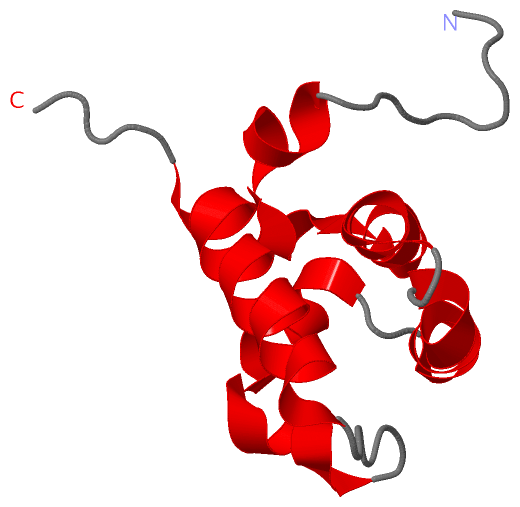
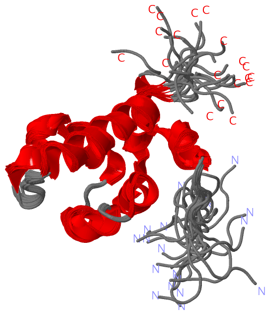
 Visualization

(-) Interactive Views

NMR Structure
  Complete Structure
    Jena3D(integrated viewing of ligand, site, SAP, PROSITE, SCOP information)
    WebMol | AstexViewer[tm]@PDBe
(Java Applets, require no local installation except for Java; loading may be slow)
    STRAP
(Java WebStart application, automatic local installation, requires Java; full application with system access!)
    RasMol
(require local installation)
    Molscript (VRML)
(requires installation of a VRML viewer; select preferred view via VRML and generate a mono or stereo PDF format file)
 
  Ligands, Modified Residues, Ions
(no "Ligands, Modified Residues, Ions" information available for 1v85)
 
  Sites
(no "Sites" information available for 1v85)
 
  Cis Peptide Bonds
    Ala A:64 - Pro A:65   [ RasMol ]  
 

(-) Still Images

Jmol
  protein: cartoon or spacefill or dots and stick; nucleic acid: cartoon and stick; ligands: spacefill; active site: stick
Molscript
  protein, nucleic acid: cartoon; ligands: spacefill; active site: ball and stick

 Databases and Analysis Tools

(-) Databases

Access by PDB/NDB ID
  1v85
    Family and Domain InformationProDom | SYSTERS
    General Structural InformationGlycoscienceDB | MMDB | NDB | OCA | PDB | PDBe | PDBj | PDBsum | PDBWiki | PQS | PROTEOPEDIA
    Orientation in MembranesOPM
    Protein SurfaceSURFACE
    Secondary StructureDSSP (structure derived) | HSSP (homology derived)
    Structural GenomicsGeneCensus
    Structural NeighboursCE | VAST
    Structure ClassificationCATH | Dali | SCOP
    Validation and Original DataBMRB Data View | BMRB Restraints Grid | EDS | PROCHECK | RECOORD | WHAT_CHECK
 
Access by UniProt ID/Accession number
  BFAR_MOUSE | Q8R079
    Comparative Protein Structure ModelsModBase
    Genomic InformationEnsembl
    Protein-protein InteractionDIP
    Sequence, Family and Domain InformationInterPro | Pfam | SMART | UniProtKB/SwissProt
 
Access by Enzyme Classificator   (EC Number)
  (no 'Enzyme Classificator' available)
    General Enzyme InformationBRENDA | EC-PDB | Enzyme | IntEnz
    PathwayKEGG | MetaCyc
 
Access by Disease Identifier   (MIM ID)
  (no 'MIM ID' available)
    Disease InformationOMIM
 
Access by GenAge ID
  (no 'GenAge ID' available)
    Age Related InformationGenAge

(-) Analysis Tools

Access by PDB/NDB ID
    Domain InformationXDom
    Interatomic Contacts of Structural UnitsCSU
    Ligand-protein ContactsLPC
    Protein CavitiescastP
    Sequence and Secondary StructurePDBCartoon
    Structure AlignmentSTRAP(Java WebStart application, automatic local installation, requires Java; full application with system access!)
    Structure and Sequence BrowserSTING
 
Access by UniProt ID/Accession number
  BFAR_MOUSE | Q8R079
    Protein Disorder PredictionDisEMBL | FoldIndex | GLOBPLOT (for more information see DisProt)

 Related Entries

(-) Entries Sharing at Least One Protein Chain (UniProt ID)

(no "Entries Sharing at Least One Protein Chain" available for 1V85)

(-) Related Entries Specified in the PDB File

(no "Related Entries Specified in the PDB File" available for 1V85)