Show PDB file:   
         Plain Text   HTML   (compressed file size)
QuickSearch:   
by PDB,NDB,UniProt,PROSITE Code or Search Term(s)  
(-)Asymmetric Unit
(-)Asym. Unit - sites
(-)Biological Unit 1
(-)Biol. Unit 1 - sites
collapse expand < >
Image Asymmetric Unit
Asymmetric Unit  (Jmol Viewer)
Image Asym. Unit - sites
Asym. Unit - sites  (Jmol Viewer)
Image Biological Unit 1
Biological Unit 1  (Jmol Viewer)
Image Biol. Unit 1 - sites
Biol. Unit 1 - sites  (Jmol Viewer)

(-) Description

Title :  RALSTONIA SOLANACEARUM LECTIN (RS-IIL) IN COMPLEX WITH ALPHA-METHYLMANNOSIDE
 
Authors :  E. P. Mitchell, M. Wimmerova, A. Imberty
Date :  22 Oct 03  (Deposition) - 28 Apr 04  (Release) - 02 Jun 09  (Revision)
Method :  X-RAY DIFFRACTION
Resolution :  1.70
Chains :  Asym. Unit :  A
Biol. Unit 1:  A  (4x)
Keywords :  Lectin, Sugar-Binding Protein, Alpha-Methyl-Mannoside (Keyword Search: [Gene Ontology, PubMed, Web (Google))
 
Reference :  D. Sudakevitz, N. Kostlanova, G. Blatman-Jan, E. P. Mitchell, B. Lerrer, M. Wimmerova, D. J. Katcoff, A. Imberty, N. Gilboa-Garber
A New Ralstonia Solanacearum High-Affinity Mannose-Binding Lectin Rs-Iil Structurally Resembling The Pseudomonas Aeruginosa Fucose-Specific Lectin Pa-Iil
Mol. Microbiol. V. 52 691 2004
PubMed-ID: 15101976  |  Reference-DOI: 10.1111/J.1365-2958.2004.04020.X

(-) Compounds

Molecule 1 - LECTIN
    Atcc11696
    ChainsA
    Organism ScientificRALSTONIA SOLANACEARUM
    Organism Taxid305
    SynonymHYPOTHETICAL PROTEIN RSC3288

 Structural Features

(-) Chains, Units

  1
Asymmetric Unit A
Biological Unit 1 (4x)A

Summary Information (see also Sequences/Alignments below)
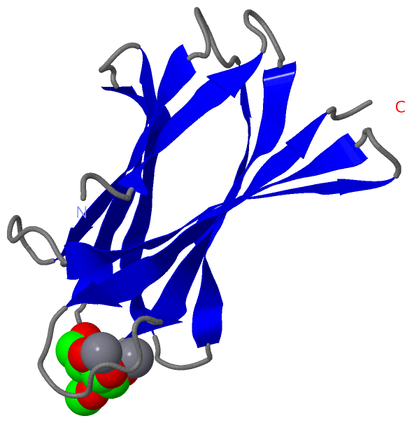
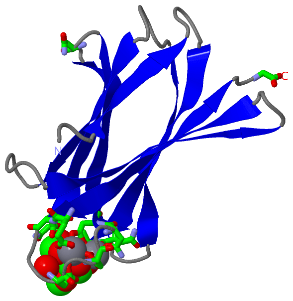
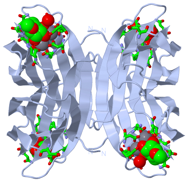
(-) Ligands, Modified Residues, Ions  (2, 3)

Asymmetric Unit (2, 3)
No.NameCountTypeFull Name
1CA2Ligand/IonCALCIUM ION
2MMA1Ligand/IonO1-METHYL-MANNOSE
Biological Unit 1 (1, 4)
No.NameCountTypeFull Name
1CA-1Ligand/IonCALCIUM ION
2MMA4Ligand/IonO1-METHYL-MANNOSE

(-) Sites  (3, 3)

Asymmetric Unit (3, 3)
No.NameEvidenceResiduesDescription
1AC1SOFTWAREASN A:21 , ALA A:22 , ALA A:23 , ASN A:24 , ASN A:34 , GLU A:94 , ASP A:95 , ASP A:98 , ASP A:100 , ASP A:103 , GLY A:113 , CA A:1114 , CA A:1115 , HOH A:2177BINDING SITE FOR RESIDUE MMA A1116
2AC2SOFTWAREASN A:21 , ASP A:100 , ASN A:102 , ASP A:103 , GLY A:113 , MMA A:1116BINDING SITE FOR RESIDUE CA A1114
3AC3SOFTWAREGLU A:94 , ASP A:98 , ASP A:100 , ASP A:103 , MMA A:1116BINDING SITE FOR RESIDUE CA A1115

(-) SS Bonds  (0, 0)

(no "SS Bond" information available for 1UQX)

(-) Cis Peptide Bonds  (1, 1)

Asymmetric Unit
No.Residues
1Trp A:110 -Pro A:111

 Sequence-Structure Mapping

(-) SAPs(SNPs)/Variants  (0, 0)

(no "SAP(SNP)/Variant" information available for 1UQX)

(-) PROSITE Motifs  (0, 0)

(no "PROSITE Motif" information available for 1UQX)

(-) Exons   (0, 0)

(no "Exon" information available for 1UQX)

(-) Sequences/Alignments

Asymmetric Unit
   Reformat: Number of residues per line =  ('0' or empty: single-line sequence representation)
  Number of residues per labelling interval =   
  UniProt sequence: complete  aligned part    
   Show mapping: SCOP domains CATH domains Pfam domains Secondary structure (by author)
SAPs(SNPs) PROSITE motifs Exons
(details for a mapped element are shown in a popup box when the mouse pointer rests over it)
Chain A from PDB  Type:PROTEIN  Length:113
 aligned with Q8XUA5_RALSO | Q8XUA5 from UniProtKB/TrEMBL  Length:114

    Alignment length:113
                                    11        21        31        41        51        61        71        81        91       101       111   
         Q8XUA5_RALSO     2 AQQGVFTLPANTSFGVTAFANAANTQTIQVLVDNVVKATFTGSGTSDKLLGSQVLNSGSGAIKIQVSVNGKPSDLVSNQTILANKLNFAMVGSEDGTDNDYNDGIAVLNWPLG 114
               SCOP domains d1uqxa_ A: Mannose-specific lectin RS-IIL                                                                         SCOP domains
               CATH domains 1uqxA00 A:1-113 Calcium-mediated lectin                                                                           CATH domains
               Pfam domains -----PA-IIL-1uqxA01 A:6-112                                                                                     - Pfam domains
         Sec.struct. author ...eeeee....eeeeeeeee...eeeeeeee...eeeeeeee...eeeeeeeee.....eeeeeee......eeeeeeeee...eeeeeeeee........eeeeeeee... Sec.struct. author
                 SAPs(SNPs) ----------------------------------------------------------------------------------------------------------------- SAPs(SNPs)
                    PROSITE ----------------------------------------------------------------------------------------------------------------- PROSITE
                 Transcript ----------------------------------------------------------------------------------------------------------------- Transcript
                 1uqx A   1 AQQGVFTLPANTSFGVTAFANAANTQTIQVLVDNVVKATFTGSGTSDKLLGSQVLNSGSGAIKIQVSVNGKPSDLVSNQTILANKLNFAMVGSEDGTDNDYNDGIAVLNWPLG 113
                                    10        20        30        40        50        60        70        80        90       100       110   

   Legend:   → Mismatch (orange background)
  - → Gap (green background, '-', border residues have a numbering label)
    → Modified Residue (blue background, lower-case, 'x' indicates undefined single-letter code, labelled with number + name)
  x → Chemical Group (purple background, 'x', labelled with number + name, e.g. ACE or NH2)
  extra numbering lines below/above indicate numbering irregularities and modified residue names etc., number ends below/above '|'

 Classification and Annotation

(-) SCOP Domains  (1, 1)

Asymmetric Unit

(-) CATH Domains  (1, 1)

Asymmetric Unit
(-)
Class: Mainly Beta (13760)

(-) Pfam Domains  (1, 1)

Asymmetric Unit

(-) Gene Ontology  (2, 2)

Asymmetric Unit(hide GO term definitions)
Chain A   (Q8XUA5_RALSO | Q8XUA5)
molecular function
    GO:0030246    carbohydrate binding    Interacting selectively and non-covalently with any carbohydrate, which includes monosaccharides, oligosaccharides and polysaccharides as well as substances derived from monosaccharides by reduction of the carbonyl group (alditols), by oxidation of one or more hydroxy groups to afford the corresponding aldehydes, ketones, or carboxylic acids, or by replacement of one or more hydroxy group(s) by a hydrogen atom. Cyclitols are generally not regarded as carbohydrates.
    GO:0046872    metal ion binding    Interacting selectively and non-covalently with any metal ion.

 Visualization

(-) Interactive Views

Asymmetric Unit
  Complete Structure
    Jena3D(integrated viewing of ligand, site, SAP, PROSITE, SCOP information)
    WebMol | AstexViewer[tm]@PDBe
(Java Applets, require no local installation except for Java; loading may be slow)
    STRAP
(Java WebStart application, automatic local installation, requires Java; full application with system access!)
    RasMol
(require local installation)
    Molscript (VRML)
(requires installation of a VRML viewer; select preferred view via VRML and generate a mono or stereo PDF format file)
 
  Ligands, Modified Residues, Ions
    CA  [ RasMol | Jena3D ]  +environment [ RasMol | Jena3D ]
    MMA  [ RasMol | Jena3D ]  +environment [ RasMol | Jena3D ]
 
  Sites
    AC1  [ RasMol ]  +environment [ RasMol ]
    AC2  [ RasMol ]  +environment [ RasMol ]
    AC3  [ RasMol ]  +environment [ RasMol ]
 
  Cis Peptide Bonds
    Trp A:110 - Pro A:111   [ RasMol ]  
 
Biological Unit
  Complete Structure
    Biological Unit 1  [ Jena3D ]

(-) Still Images

Jmol
  protein: cartoon or spacefill or dots and stick; nucleic acid: cartoon and stick; ligands: spacefill; active site: stick
Molscript
  protein, nucleic acid: cartoon; ligands: spacefill; active site: ball and stick

 Databases and Analysis Tools

(-) Databases

Access by PDB/NDB ID
  1uqx
    Family and Domain InformationProDom | SYSTERS
    General Structural InformationGlycoscienceDB | MMDB | NDB | OCA | PDB | PDBe | PDBj | PDBsum | PDBWiki | PQS | PROTEOPEDIA
    Orientation in MembranesOPM
    Protein SurfaceSURFACE
    Secondary StructureDSSP (structure derived) | HSSP (homology derived)
    Structural GenomicsGeneCensus
    Structural NeighboursCE | VAST
    Structure ClassificationCATH | Dali | SCOP
    Validation and Original DataBMRB Data View | BMRB Restraints Grid | EDS | PROCHECK | RECOORD | WHAT_CHECK
 
Access by UniProt ID/Accession number
  Q8XUA5_RALSO | Q8XUA5
    Comparative Protein Structure ModelsModBase
    Genomic InformationEnsembl
    Protein-protein InteractionDIP
    Sequence, Family and Domain InformationInterPro | Pfam | SMART | UniProtKB/TrEMBL
 
Access by Enzyme Classificator   (EC Number)
  (no 'Enzyme Classificator' available)
    General Enzyme InformationBRENDA | EC-PDB | Enzyme | IntEnz
    PathwayKEGG | MetaCyc
 
Access by Disease Identifier   (MIM ID)
  (no 'MIM ID' available)
    Disease InformationOMIM
 
Access by GenAge ID
  (no 'GenAge ID' available)
    Age Related InformationGenAge

(-) Analysis Tools

Access by PDB/NDB ID
    Domain InformationXDom
    Interatomic Contacts of Structural UnitsCSU
    Ligand-protein ContactsLPC
    Protein CavitiescastP
    Sequence and Secondary StructurePDBCartoon
    Structure AlignmentSTRAP(Java WebStart application, automatic local installation, requires Java; full application with system access!)
    Structure and Sequence BrowserSTING
 
Access by UniProt ID/Accession number
  Q8XUA5_RALSO | Q8XUA5
    Protein Disorder PredictionDisEMBL | FoldIndex | GLOBPLOT (for more information see DisProt)

 Related Entries

(-) Entries Sharing at Least One Protein Chain (UniProt ID)

UniProtKB/TrEMBL
        Q8XUA5_RALSO | Q8XUA52chh

(-) Related Entries Specified in the PDB File

(no "Related Entries Specified in the PDB File" available for 1UQX)