|
|
|
|
 Description
|
|
Compounds
|
||||||||||||||||||||||||||||||||||||||||||||||||||||
Chains, Units
Summary Information (see also Sequences/Alignments below) |
Ligands, Modified Residues, Ions (0, 0)| (no "Ligand,Modified Residues,Ions" information available for 1UL7) |
Sites (0, 0)| (no "Site" information available for 1UL7) |
SS Bonds (0, 0)| (no "SS Bond" information available for 1UL7) |
Cis Peptide Bonds (0, 0)| (no "Cis Peptide Bond" information available for 1UL7) |
SAPs(SNPs)/Variants (0, 0)| (no "SAP(SNP)/Variant" information available for 1UL7) |
PROSITE Motifs (1, 1)
NMR Structure (1, 1)
|
||||||||||||||||||||||||
Exons (0, 0)| (no "Exon" information available for 1UL7) |
Sequences/Alignments
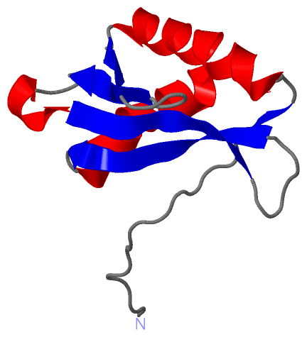
NMR StructureChain A from PDB Type:PROTEIN Length:102 aligned with MARK3_MOUSE | Q03141 from UniProtKB/Swiss-Prot Length:753 Alignment length:154 609 619 629 639 649 659 669 679 689 699 709 719 729 739 749 MARK3_MOUSE 600 GSTNLFSKLTSKLTRRNMSFRFIKRLPTEYERNGRYEGSSRNVSSEQKDENREAKPRSLRFTWSMKTTSSMDPSDMMREIRKVLDANNCDYEQRERFLLFCVHGDGHAENLVQWEMEVCKLPRLSLNGVRFKRISGTSIAFKNIASKIANELKL 753 SCOP domains d1u l7a_ A: Map/microtubule affinity-regulating kinase 3 SCOP domains CATH domains 1ul 7A00 A:1-102 Kinase associated domain 1, KA1 CATH domains Pfam domains -----------------------------------------------------------------------------------------------------------KA1-1ul7A01 A:56-102 Pfam domains SAPs(SNPs) ---------------------------------------------------------------------------------------------------------------------------------------------------------- SAPs(SNPs) PROSITE --------------------------------------------------------------------------------------------------------KA1 PDB: A:53-102 UniProt: 704-753 PROSITE Transcript ---------------------------------------------------------------------------------------------------------------------------------------------------------- Transcript 1ul7 A 1 GSS----------------------------------GSSG------------------RFTWSMKTTSSMDPSDMMREIRKVLGANNCDYEQRERFLLFCVHGDGHAENLVQWEMEVCKLPRLSLNGVRFKRISGTSIAFKNIASKIANELKL 102 | - - - | 6| - 8 18 28 38 48 58 68 78 88 98 3 4 7 8
|
||||||||||||||||||||
SCOP Domains (1, 1)
NMR Structure
 |
CATH Domains (1, 1)
NMR Structure
|
Pfam Domains (1, 1)
NMR Structure
|
Gene Ontology (11, 11)|
NMR Structure(hide GO term definitions) Chain A (MARK3_MOUSE | Q03141)
|
||||||||||||||||||||||||||||||||||||||||||||||||||||||||||||||||||||||||||||||||||||
Interactive Views
|
||||||||||||||||||||||||||||||||||||||||||||||||||||||||||||||||||||||||||||||||||||||||||||||||||||||||||||||||||||
Still Images
|
||||||||||||||||
Databases
|
||||||||||||||||||||||||||||||||||||||||||||||||||||||||||||||||||||||||||||||||||||||||||||||||||||||||||||||||||||||||||||||||||||||||||||||||||||||||||||||||
Analysis Tools
|
|||||||||||||||||||||||||||||||||||||||||||||||||||||||||||||
Entries Sharing at Least One Protein Chain (UniProt ID)
Related Entries Specified in the PDB File
|
|