| molecular function |
|---|
| | GO:0016787 | | hydrolase activity | | Catalysis of the hydrolysis of various bonds, e.g. C-O, C-N, C-C, phosphoric anhydride bonds, etc. Hydrolase is the systematic name for any enzyme of EC class 3. |
| | GO:0008969 | | phosphohistidine phosphatase activity | | Catalysis of the reaction: phosphohistidine + H2O = histidine + phosphate. |
| | GO:0004721 | | phosphoprotein phosphatase activity | | Catalysis of the reaction: a phosphoprotein + H2O = a protein + phosphate. Together with protein kinases, these enzymes control the state of phosphorylation of cell proteins and thereby provide an important mechanism for regulating cellular activity. |
| | GO:0005515 | | protein binding | | Interacting selectively and non-covalently with any protein or protein complex (a complex of two or more proteins that may include other nonprotein molecules). |
| biological process |
|---|
| | GO:0006464 | | cellular protein modification process | | The covalent alteration of one or more amino acids occurring in proteins, peptides and nascent polypeptides (co-translational, post-translational modifications) occurring at the level of an individual cell. Includes the modification of charged tRNAs that are destined to occur in a protein (pre-translation modification). |
| | GO:0016311 | | dephosphorylation | | The process of removing one or more phosphoric (ester or anhydride) residues from a molecule. |
| | GO:0006470 | | protein dephosphorylation | | The process of removing one or more phosphoric residues from a protein. |
| cellular component |
|---|
| | GO:0005737 | | cytoplasm | | All of the contents of a cell excluding the plasma membrane and nucleus, but including other subcellular structures. |
| | GO:0005829 | | cytosol | | The part of the cytoplasm that does not contain organelles but which does contain other particulate matter, such as protein complexes. |
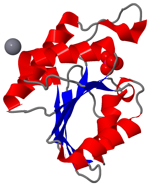
 Description
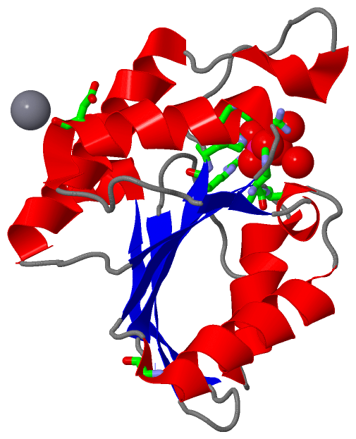