|
|
|
|
 Description
|
|
Compounds
|
||||||||||||||||||||||||||||||||||||||||||||||||
Chains, Units
Summary Information (see also Sequences/Alignments below) |
Ligands, Modified Residues, Ions (0, 0)| (no "Ligand,Modified Residues,Ions" information available for 1UG7) |
Sites (0, 0)| (no "Site" information available for 1UG7) |
SS Bonds (0, 0)| (no "SS Bond" information available for 1UG7) |
Cis Peptide Bonds (0, 0)| (no "Cis Peptide Bond" information available for 1UG7) |
SAPs(SNPs)/Variants (0, 0)| (no "SAP(SNP)/Variant" information available for 1UG7) |
PROSITE Motifs (0, 0)| (no "PROSITE Motif" information available for 1UG7) |
Exons (0, 0)| (no "Exon" information available for 1UG7) |
Sequences/Alignments
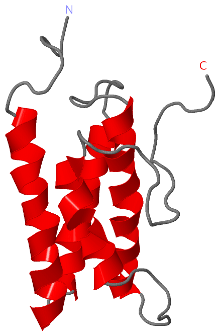
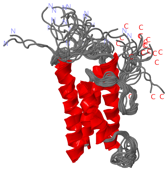
NMR StructureChain A from PDB Type:PROTEIN Length:128 aligned with AIDA_MOUSE | Q8C4Q6 from UniProtKB/Swiss-Prot Length:305 Alignment length:149 1 | 3 13 23 33 43 53 63 73 83 93 103 113 123 133 AIDA_MOUSE - -------MSEVTRSLLQRWGASLRRGADFDSWGQLVEAIDEYQILARHLQKEAQAQHNNSEFTEEQKKTIGKIATCLELRSAALQSTQSQEEFKLEDLKKLEPILKNILTYNKEFPFDVQPIPLRRILAPGEEENLEFEEDEEGGAGAG 142 SCOP domains d1ug7a_ A: Domain from hypothetical 2610208m17rik protein SCOP domains CATH domains 1ug7A00 A:1-128 Domain from hypothetical 2610208m17rik protein CATH domains Pfam domains --------------Aida-C2-1ug7A01 A:15-120 ----------------------------- Pfam domains SAPs(SNPs) ----------------------------------------------------------------------------------------------------------------------------------------------------- SAPs(SNPs) PROSITE ----------------------------------------------------------------------------------------------------------------------------------------------------- PROSITE Transcript ----------------------------------------------------------------------------------------------------------------------------------------------------- Transcript 1ug7 A 1 GSSGSSGMSEVTRSLLQRWGASLRRGADFDSWGQLVEAIDEYQILARHLQKEAQAQHNNSEFTEEQKKTIGKIATCLELRSAALQSTQSQEEFKLEDLKKLEPILKNILTYNKEFPFDVQPIS---------------------GPSSG 128 10 20 30 40 50 60 70 80 90 100 110 120 | - - | 123 124
|
||||||||||||||||||||
SCOP Domains (1, 1)
NMR Structure
 |
CATH Domains (1, 1)
NMR Structure
|
Pfam Domains (1, 1)
NMR Structure
|
Gene Ontology (11, 11)|
NMR Structure(hide GO term definitions) Chain A (AIDA_MOUSE | Q8C4Q6)
|
||||||||||||||||||||||||||||||||||||||||||||||||||||||||||||||||||||||||||||||||||||
Interactive Views
|
||||||||||||||||||||||||||||||||||||||||||||||||||||||||||||||||||||||||||||||||||||||||||||||||||||||||||||||||||||
Still Images
|
||||||||||||||||
Databases
|
||||||||||||||||||||||||||||||||||||||||||||||||||||||||||||||||||||||||||||||||||||||||||||||||||||||||||||||||||||||||||||||||||||||||||||||||||||||||||||||||
Analysis Tools
|
|||||||||||||||||||||||||||||||||||||||||||||||||||||||||||||
Entries Sharing at Least One Protein Chain (UniProt ID)
Related Entries Specified in the PDB File
|
|