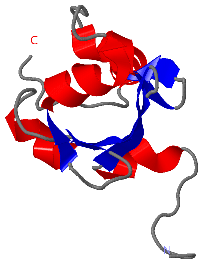
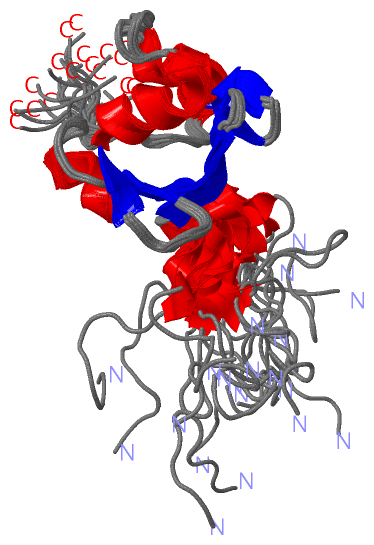
Show PDB file:   
         Plain Text   HTML   (compressed file size)
QuickSearch:   
by PDB,NDB,UniProt,PROSITE Code or Search Term(s)  
(-)NMR Structure - model 1
(-)NMR Structure - all models
collapse expand < >
Image NMR Structure - model 1
NMR Structure - model 1  (Jmol Viewer)
Image NMR Structure - all models
NMR Structure - all models  (Jmol Viewer)

(-) Description

Title :  SOLUTION STRUCTURE OF THE SAND DOMAIN OF THE PUTATIVE NUCLEAR PROTEIN HOMOLOG (5830484A20RIK)
 
Authors :  N. Tochio, N. Kobayashi, S. Koshiba, T. Kigawa, M. Inoue, M. Shirouzu, T. Terada, T. Yabuki, M. Aoki, E. Seki, T. Matsuda, H. Hirota, M. Yoshida, A. Tanaka, T. Osanai, Y. Matsuo, T. Arakawa, P. Carninci, J. Kawai, Y. Hayashizaki, S. Yokoyama, Riken Structural Genomics/Proteomics Initiative (Rsgi)
Date :  02 Jun 03  (Deposition) - 22 Jun 04  (Release) - 24 Feb 09  (Revision)
Method :  SOLUTION NMR
Resolution :  NOT APPLICABLE
Chains :  NMR Structure  :  A  (20x)
Keywords :  Sand Domain, Kdwk Motif, Nuclear Protein, Structural Genomics, Riken Structural Genomics/Proteomics Initiative, Rsgi, Unknown Function (Keyword Search: [Gene Ontology, PubMed, Web (Google))
 
Reference :  N. Tochio, N. Kobayashi, S. Koshiba, T. Kigawa, M. Inoue, M. Shirouzu, T. Terada, T. Yabuki, M. Aoki, E. Seki, T. Matsuda, H. Hirota, M. Yoshida, A. Tanaka, T. Osanai, Y. Matsuo, T. Arakawa, P. Carninci, J. Kawai, Y. Hayashizaki, S. Yokoyama
Solution Structure Of The Sand Domain Of The Putative Nuclear Protein Homolog (5830484A20Rik)
To Be Published
PubMed: search
(for further references see the PDB file header)

(-) Compounds

Molecule 1 - PUTATIVE NUCLEAR PROTEIN HOMOLOG 5830484A20RIK
    ChainsA
    EngineeredYES
    Expression System PlasmidP021030-59
    Expression System Vector TypePLASMID
    FragmentSAND DOMAIN
    GeneRIKEN CDNA 5830484A20
    Organism CommonHOUSE MOUSE
    Organism ScientificMUS MUSCULUS
    Organism Taxid10090
    Other DetailsCELL-FREE PROTEIN SYNTHESIS

 Structural Features

(-) Chains, Units

  
NMR Structure (20x)

Summary Information (see also Sequences/Alignments below)

(-) Ligands, Modified Residues, Ions  (0, 0)

(no "Ligand,Modified Residues,Ions" information available for 1UFN)

(-) Sites  (0, 0)

(no "Site" information available for 1UFN)

(-) SS Bonds  (0, 0)

(no "SS Bond" information available for 1UFN)

(-) Cis Peptide Bonds  (0, 0)

(no "Cis Peptide Bond" information available for 1UFN)

 Sequence-Structure Mapping

(-) SAPs(SNPs)/Variants  (0, 0)

(no "SAP(SNP)/Variant" information available for 1UFN)

(-) PROSITE Motifs  (1, 1)

NMR Structure (1, 1)
 PROSITEUniProtKBPDB
No.IDACDescriptionIDLocationCountLocation
1SANDPS50864 SAND domain profile.SP110_MOUSE353-433  1A:8-88

(-) Exons   (0, 0)

(no "Exon" information available for 1UFN)

(-) Sequences/Alignments

NMR Structure
   Reformat: Number of residues per line =  ('0' or empty: single-line sequence representation)
  Number of residues per labelling interval =   
  UniProt sequence: complete  aligned part    
   Show mapping: SCOP domains CATH domains Pfam domains Secondary structure (by author)
SAPs(SNPs) PROSITE motifs Exons
(details for a mapped element are shown in a popup box when the mouse pointer rests over it)
Chain A from PDB  Type:PROTEIN  Length:94
 aligned with SP110_MOUSE | Q8BVK9 from UniProtKB/Swiss-Prot  Length:445

    Alignment length:117
                                   337       347       357       367       377       387       397       407       417       427       437       
          SP110_MOUSE   328 EASKTSGRKRPSTARRTTQVPEKTKNDAVDFSPTLPVTCGKAKGTLFQEKLKQGASKKCIQNEAGDWLTVKEFLNEGGRATSKDWKGVIRCNGETLRHLEQKGLLFFTSKSKPQKKG 444
               SCOP domains d1ufna_                   A: Putative nuclear protein homolog 5830484a20rik                                           SCOP domains
               CATH domains 1ufnA00                   A:1-94 SAND domain                                                                          CATH domains
               Pfam domains -------------------------SAND-1ufnA01 A:8-88                                                              ----------- Pfam domains
         Sec.struct. author .......------------------.hhhhhh.eeeeee..eeeeeehhhhhh......ee.....eehhhhhhhhhh.....hhhhhhee..eehhhhhhh.........-----. Sec.struct. author
                 SAPs(SNPs) --------------------------------------------------------------------------------------------------------------------- SAPs(SNPs)
                    PROSITE -------------------------SAND  PDB: A:8-88 UniProt: 353-433                                               ----------- PROSITE
                 Transcript --------------------------------------------------------------------------------------------------------------------- Transcript
                 1ufn A   1 GSSGSSG------------------NDAVDFSPTLPVTCGKAKGTLFQEKLKQGASKKCIQNEAGDWLTVKEFLNEGGRATSKDWKGVIRCNGETLRHLEQKGLLFSGPSS-----G  94
                                  |  -         -     |  12        22        32        42        52        62        72        82        92|     |
                                  7                  8                                                                                   93    94

   Legend:   → Mismatch (orange background)
  - → Gap (green background, '-', border residues have a numbering label)
    → Modified Residue (blue background, lower-case, 'x' indicates undefined single-letter code, labelled with number + name)
  x → Chemical Group (purple background, 'x', labelled with number + name, e.g. ACE or NH2)
  extra numbering lines below/above indicate numbering irregularities and modified residue names etc., number ends below/above '|'

 Classification and Annotation

(-) SCOP Domains  (1, 1)

NMR Structure

(-) CATH Domains  (1, 1)

NMR Structure
(-)
Class: Alpha Beta (26913)

(-) Pfam Domains  (1, 1)

NMR Structure

(-) Gene Ontology  (10, 10)

NMR Structure(hide GO term definitions)
Chain A   (SP110_MOUSE | Q8BVK9)
molecular function
    GO:0003677    DNA binding    Any molecular function by which a gene product interacts selectively and non-covalently with DNA (deoxyribonucleic acid).
biological process
    GO:0006915    apoptotic process    A programmed cell death process which begins when a cell receives an internal (e.g. DNA damage) or external signal (e.g. an extracellular death ligand), and proceeds through a series of biochemical events (signaling pathway phase) which trigger an execution phase. The execution phase is the last step of an apoptotic process, and is typically characterized by rounding-up of the cell, retraction of pseudopodes, reduction of cellular volume (pyknosis), chromatin condensation, nuclear fragmentation (karyorrhexis), plasma membrane blebbing and fragmentation of the cell into apoptotic bodies. When the execution phase is completed, the cell has died.
    GO:0002376    immune system process    Any process involved in the development or functioning of the immune system, an organismal system for calibrated responses to potential internal or invasive threats.
    GO:0045087    innate immune response    Innate immune responses are defense responses mediated by germline encoded components that directly recognize components of potential pathogens.
    GO:0043065    positive regulation of apoptotic process    Any process that activates or increases the frequency, rate or extent of cell death by apoptotic process.
    GO:0006355    regulation of transcription, DNA-templated    Any process that modulates the frequency, rate or extent of cellular DNA-templated transcription.
    GO:0009617    response to bacterium    Any process that results in a change in state or activity of a cell or an organism (in terms of movement, secretion, enzyme production, gene expression, etc.) as a result of a stimulus from a bacterium.
    GO:0006351    transcription, DNA-templated    The cellular synthesis of RNA on a template of DNA.
cellular component
    GO:0005730    nucleolus    A small, dense body one or more of which are present in the nucleus of eukaryotic cells. It is rich in RNA and protein, is not bounded by a limiting membrane, and is not seen during mitosis. Its prime function is the transcription of the nucleolar DNA into 45S ribosomal-precursor RNA, the processing of this RNA into 5.8S, 18S, and 28S components of ribosomal RNA, and the association of these components with 5S RNA and proteins synthesized outside the nucleolus. This association results in the formation of ribonucleoprotein precursors; these pass into the cytoplasm and mature into the 40S and 60S subunits of the ribosome.
    GO:0005634    nucleus    A membrane-bounded organelle of eukaryotic cells in which chromosomes are housed and replicated. In most cells, the nucleus contains all of the cell's chromosomes except the organellar chromosomes, and is the site of RNA synthesis and processing. In some species, or in specialized cell types, RNA metabolism or DNA replication may be absent.

 Visualization

(-) Interactive Views

NMR Structure
  Complete Structure
    Jena3D(integrated viewing of ligand, site, SAP, PROSITE, SCOP information)
    WebMol | AstexViewer[tm]@PDBe
(Java Applets, require no local installation except for Java; loading may be slow)
    STRAP
(Java WebStart application, automatic local installation, requires Java; full application with system access!)
    RasMol
(require local installation)
    Molscript (VRML)
(requires installation of a VRML viewer; select preferred view via VRML and generate a mono or stereo PDF format file)
 
  Ligands, Modified Residues, Ions
(no "Ligands, Modified Residues, Ions" information available for 1ufn)
 
  Sites
(no "Sites" information available for 1ufn)
 
  Cis Peptide Bonds
(no "Cis Peptide Bonds" information available for 1ufn)
 

(-) Still Images

Jmol
  protein: cartoon or spacefill or dots and stick; nucleic acid: cartoon and stick; ligands: spacefill; active site: stick
Molscript
  protein, nucleic acid: cartoon; ligands: spacefill; active site: ball and stick

 Databases and Analysis Tools

(-) Databases

Access by PDB/NDB ID
  1ufn
    Family and Domain InformationProDom | SYSTERS
    General Structural InformationGlycoscienceDB | MMDB | NDB | OCA | PDB | PDBe | PDBj | PDBsum | PDBWiki | PQS | PROTEOPEDIA
    Orientation in MembranesOPM
    Protein SurfaceSURFACE
    Secondary StructureDSSP (structure derived) | HSSP (homology derived)
    Structural GenomicsGeneCensus
    Structural NeighboursCE | VAST
    Structure ClassificationCATH | Dali | SCOP
    Validation and Original DataBMRB Data View | BMRB Restraints Grid | EDS | PROCHECK | RECOORD | WHAT_CHECK
 
Access by UniProt ID/Accession number
  SP110_MOUSE | Q8BVK9
    Comparative Protein Structure ModelsModBase
    Genomic InformationEnsembl
    Protein-protein InteractionDIP
    Sequence, Family and Domain InformationInterPro | Pfam | SMART | UniProtKB/SwissProt
 
Access by Enzyme Classificator   (EC Number)
  (no 'Enzyme Classificator' available)
    General Enzyme InformationBRENDA | EC-PDB | Enzyme | IntEnz
    PathwayKEGG | MetaCyc
 
Access by Disease Identifier   (MIM ID)
  (no 'MIM ID' available)
    Disease InformationOMIM
 
Access by GenAge ID
  (no 'GenAge ID' available)
    Age Related InformationGenAge

(-) Analysis Tools

Access by PDB/NDB ID
    Domain InformationXDom
    Interatomic Contacts of Structural UnitsCSU
    Ligand-protein ContactsLPC
    Protein CavitiescastP
    Sequence and Secondary StructurePDBCartoon
    Structure AlignmentSTRAP(Java WebStart application, automatic local installation, requires Java; full application with system access!)
    Structure and Sequence BrowserSTING
 
Access by UniProt ID/Accession number
  SP110_MOUSE | Q8BVK9
    Protein Disorder PredictionDisEMBL | FoldIndex | GLOBPLOT (for more information see DisProt)

 Related Entries

(-) Entries Sharing at Least One Protein Chain (UniProt ID)

(no "Entries Sharing at Least One Protein Chain" available for 1UFN)

(-) Related Entries Specified in the PDB File

(no "Related Entries Specified in the PDB File" available for 1UFN)