|
|
|
|
 Description
|
|
Compounds
|
||||||||||||||||||||||||||||||||||||||||
Chains, Units
Summary Information (see also Sequences/Alignments below) |
Ligands, Modified Residues, Ions (1, 1)
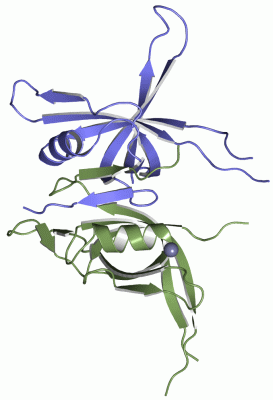
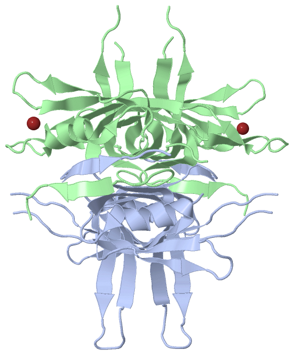
Asymmetric Unit (1, 1)
|
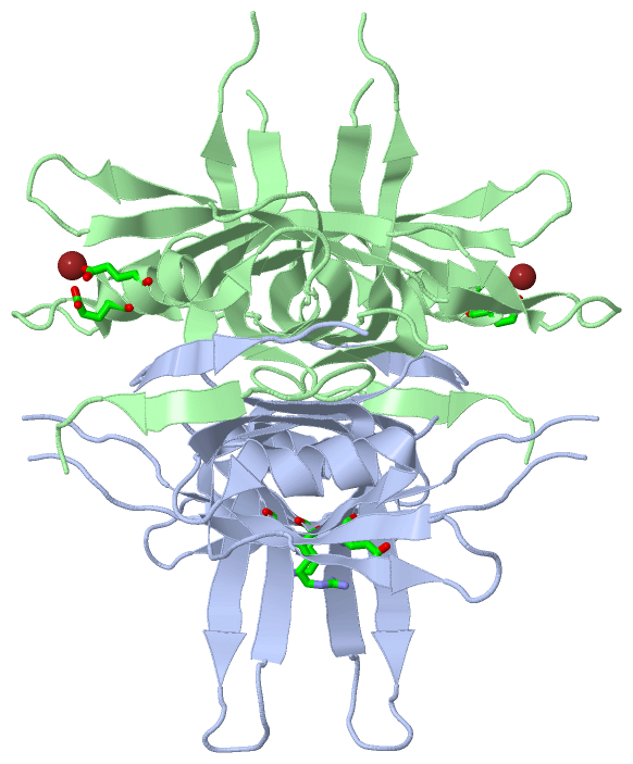
Sites (1, 1)
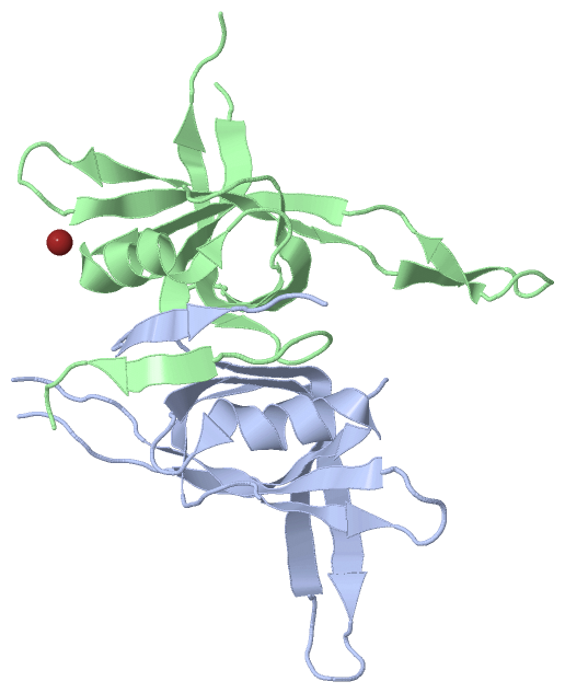
Asymmetric Unit (1, 1)
|
SS Bonds (0, 0)| (no "SS Bond" information available for 1UE1) |
Cis Peptide Bonds (0, 0)| (no "Cis Peptide Bond" information available for 1UE1) |
SAPs(SNPs)/Variants (0, 0)| (no "SAP(SNP)/Variant" information available for 1UE1) |
PROSITE Motifs (0, 0)| (no "PROSITE Motif" information available for 1UE1) |
Exons (0, 0)| (no "Exon" information available for 1UE1) |
Sequences/Alignments
Asymmetric UnitChain A from PDB Type:PROTEIN Length:113 aligned with SSB_MYCTO | P9WGD4 from UniProtKB/Swiss-Prot Length:164 Alignment length:118 12 22 32 42 52 62 72 82 92 102 112 SSB_MYCTO 3 GDTTITIVGNLTADPELRFTPSGAAVANFTVASTPRIYDRQTGEWKDGEALFLRCNIWREAAENVAESLTRGARVIVSGRLKQRSFETREGEKRTVIEVEVDEIGPSLRYATAKVNKA 120 SCOP domains d1ue1a_ A: ssDNA-binding protein SCOP domains CATH domains 1ue1A00 A:3-120 Nucleic acid-binding pr oteins CATH domains Pfam domains ---------------------------------------------------------------------------------------------------------------------- Pfam domains SAPs(SNPs) ---------------------------------------------------------------------------------------------------------------------- SAPs(SNPs) PROSITE ---------------------------------------------------------------------------------------------------------------------- PROSITE Transcript ---------------------------------------------------------------------------------------------------------------------- Transcript 1ue1 A 3 GDTTITIVGNLTADPELRFTPSGAAVANFTVASTPRIYD-----WKDGEALFLRCNIWREAAENVAESLTRGARVIVSGRLKQRSFETREGEKRTVIEVEVDEIGPSLRYATAKVNKA 120 12 22 32 |- | 52 62 72 82 92 102 112 41 47 Chain A from PDB Type:PROTEIN Length:113 aligned with SSB_MYCTU | P9WGD5 from UniProtKB/Swiss-Prot Length:164 Alignment length:118 12 22 32 42 52 62 72 82 92 102 112 SSB_MYCTU 3 GDTTITIVGNLTADPELRFTPSGAAVANFTVASTPRIYDRQTGEWKDGEALFLRCNIWREAAENVAESLTRGARVIVSGRLKQRSFETREGEKRTVIEVEVDEIGPSLRYATAKVNKA 120 SCOP domains d1ue1a_ A: ssDNA-binding protein SCOP domains CATH domains 1ue1A00 A:3-120 Nucleic acid-binding pr oteins CATH domains Pfam domains ---------------------------------------------------------------------------------------------------------------------- Pfam domains SAPs(SNPs) ---------------------------------------------------------------------------------------------------------------------- SAPs(SNPs) PROSITE ---------------------------------------------------------------------------------------------------------------------- PROSITE Transcript ---------------------------------------------------------------------------------------------------------------------- Transcript 1ue1 A 3 GDTTITIVGNLTADPELRFTPSGAAVANFTVASTPRIYD-----WKDGEALFLRCNIWREAAENVAESLTRGARVIVSGRLKQRSFETREGEKRTVIEVEVDEIGPSLRYATAKVNKA 120 12 22 32 |- | 52 62 72 82 92 102 112 41 47 Chain B from PDB Type:PROTEIN Length:119 aligned with SSB_MYCTO | P9WGD4 from UniProtKB/Swiss-Prot Length:164 Alignment length:122 11 21 31 41 51 61 71 81 91 101 111 121 SSB_MYCTO 2 AGDTTITIVGNLTADPELRFTPSGAAVANFTVASTPRIYDRQTGEWKDGEALFLRCNIWREAAENVAESLTRGARVIVSGRLKQRSFETREGEKRTVIEVEVDEIGPSLRYATAKVNKASRS 123 SCOP domains d1ue1b_ B: ssDNA-binding protein SCOP domains CATH domains 1ue1B00 B:2-123 Nucleic acid-binding proteins CATH domains Pfam domains (1) --SSB-1ue1B01 B:4-109 -------------- Pfam domains (1) Pfam domains (2) --SSB-1ue1B02 B:4-109 -------------- Pfam domains (2) SAPs(SNPs) -------------------------------------------------------------------------------------------------------------------------- SAPs(SNPs) PROSITE -------------------------------------------------------------------------------------------------------------------------- PROSITE Transcript -------------------------------------------------------------------------------------------------------------------------- Transcript 1ue1 B 2 AGDTTITIVGNLTADPELRFTPSGAAVANFTVASTPRIYDRQTGEWKDGEALFLRCNIWREAAENVAESLTRGARVIVSGRLKQRSFETR---KRTVIEVEVDEIGPSLRYATAKVNKASRS 123 11 21 31 41 51 61 71 81 91 | 101 111 121 91 95 Chain B from PDB Type:PROTEIN Length:119 aligned with SSB_MYCTU | P9WGD5 from UniProtKB/Swiss-Prot Length:164 Alignment length:122 11 21 31 41 51 61 71 81 91 101 111 121 SSB_MYCTU 2 AGDTTITIVGNLTADPELRFTPSGAAVANFTVASTPRIYDRQTGEWKDGEALFLRCNIWREAAENVAESLTRGARVIVSGRLKQRSFETREGEKRTVIEVEVDEIGPSLRYATAKVNKASRS 123 SCOP domains d1ue1b_ B: ssDNA-binding protein SCOP domains CATH domains 1ue1B00 B:2-123 Nucleic acid-binding proteins CATH domains Pfam domains (1) --SSB-1ue1B01 B:4-109 -------------- Pfam domains (1) Pfam domains (2) --SSB-1ue1B02 B:4-109 -------------- Pfam domains (2) SAPs(SNPs) -------------------------------------------------------------------------------------------------------------------------- SAPs(SNPs) PROSITE -------------------------------------------------------------------------------------------------------------------------- PROSITE Transcript -------------------------------------------------------------------------------------------------------------------------- Transcript 1ue1 B 2 AGDTTITIVGNLTADPELRFTPSGAAVANFTVASTPRIYDRQTGEWKDGEALFLRCNIWREAAENVAESLTRGARVIVSGRLKQRSFETR---KRTVIEVEVDEIGPSLRYATAKVNKASRS 123 11 21 31 41 51 61 71 81 91 | 101 111 121 91 95
|
||||||||||||||||||||
SCOP Domains (1, 2)
Asymmetric Unit
 |
CATH Domains (1, 2)
Asymmetric Unit
|
Pfam Domains (1, 2)
Asymmetric Unit
|
Gene Ontology (8, 11)|
Asymmetric Unit(hide GO term definitions) Chain A,B (SSB_MYCTU | P9WGD5)
Chain A,B (SSB_MYCTO | P9WGD4)
|
||||||||||||||||||||||||||||||||||||||||||||||||||||||||||||||||||||||||||||||||||||||||||||||||
Interactive Views
|
||||||||||||||||||||||||||||||||||||||||||||||||||||||||||||||||||||||||||||||||||||||||||||||||||||||||||||||||||||||||||||||||||||||||
Still Images
|
||||||||||||||||
Databases
|
||||||||||||||||||||||||||||||||||||||||||||||||||||||||||||||||||||||||||||||||||||||||||||||||||||||||||||||||||||||||||||||||||||||||||||||||||||||||||||||||||||||||||||||||||||||||||
Analysis Tools
|
||||||||||||||||||||||||||||||||||||||||||||||||||||||||||||||||||||||||
Entries Sharing at Least One Protein Chain (UniProt ID)
Related Entries Specified in the PDB File
|
|