Show PDB file:   
         Plain Text   HTML   (compressed file size)
QuickSearch:   
by PDB,NDB,UniProt,PROSITE Code or Search Term(s)  
(-)Asym./Biol. Unit
(-)Asym./Biol. Unit - sites
collapse expand < >
Image Asym./Biol. Unit
Asym./Biol. Unit  (Jmol Viewer)
Image Asym./Biol. Unit - sites
Asym./Biol. Unit - sites  (Jmol Viewer)

(-) Description

Title :  CRYSTAL STRUCTURE OF AMYK38 WITH LITHIUM ION
 
Authors :  T. Nonaka, M. Fujihashi, A. Kita, H. Hagihara, K. Ozaki, S. Ito, K. Miki
Date :  28 Apr 03  (Deposition) - 22 Jul 03  (Release) - 24 Feb 09  (Revision)
Method :  X-RAY DIFFRACTION
Resolution :  2.88
Chains :  Asym./Biol. Unit :  A
Keywords :  Calcium-Free, Alkaline, Alpha-Amylase, Hydrolase (Keyword Search: [Gene Ontology, PubMed, Web (Google))
 
Reference :  T. Nonaka, M. Fujihashi, A. Kita, H. Hagihara, K. Ozaki, S. Ito, K. Miki
Crystal Structure Of Calcium-Free Alpha-Amylase From Bacillus Sp. Strain Ksm-K38 (Amyk38) And Its Sodium Ion Binding Sites
J. Biol. Chem. V. 278 24818 2003
PubMed-ID: 12719434  |  Reference-DOI: 10.1074/JBC.M212763200
(for further references see the PDB file header)

(-) Compounds

Molecule 1 - AMYLASE
    ChainsA
    EC Number3.2.1.1
    EngineeredYES
    Expression SystemBACILLUS SUBTILIS
    Expression System PlasmidPHSP64
    Expression System Taxid1423
    Expression System Vector TypePLASMID
    FragmentRESIDUES 1-480
    Organism ScientificBACILLUS SP. KSM-K38
    Organism Taxid129736
    SynonymALPHA-AMYLASE

 Structural Features

(-) Chains, Units

  1
Asymmetric/Biological Unit A

Summary Information (see also Sequences/Alignments below)

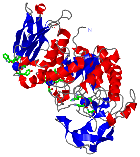
(-) Ligands, Modified Residues, Ions  (1, 3)

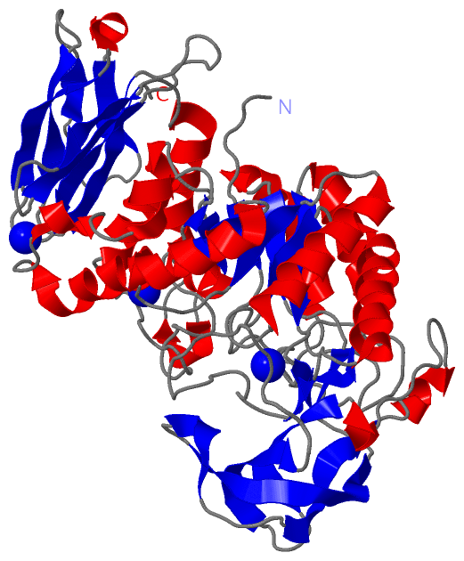
Asymmetric/Biological Unit (1, 3)
No.NameCountTypeFull Name
1NA3Ligand/IonSODIUM ION

(-) Sites  (3, 3)

Asymmetric Unit (3, 3)
No.NameEvidenceResiduesDescription
1AC1SOFTWAREASN A:104 , ASP A:194 , ASN A:200 , HIS A:235BINDING SITE FOR RESIDUE NA A 1001
2AC2SOFTWAREGLY A:300 , TYR A:302 , TRP A:403 , ASP A:404 , ASN A:427 , HOH A:2018BINDING SITE FOR RESIDUE NA A 1002
3AC3SOFTWAREASN A:289 , VAL A:324 , ASP A:325 , SER A:337 , VAL A:339BINDING SITE FOR RESIDUE NA A 1003

(-) SS Bonds  (0, 0)

(no "SS Bond" information available for 1UD8)

(-) Cis Peptide Bonds  (1, 1)

Asymmetric/Biological Unit
No.Residues
1Trp A:184 -Arg A:185

 Sequence-Structure Mapping

(-) SAPs(SNPs)/Variants  (0, 0)

(no "SAP(SNP)/Variant" information available for 1UD8)

(-) PROSITE Motifs  (0, 0)

(no "PROSITE Motif" information available for 1UD8)

(-) Exons   (0, 0)

(no "Exon" information available for 1UD8)

(-) Sequences/Alignments

Asymmetric/Biological Unit
   Reformat: Number of residues per line =  ('0' or empty: single-line sequence representation)
  Number of residues per labelling interval =   
  UniProt sequence: complete  aligned part    
   Show mapping: SCOP domains CATH domains Pfam domains Secondary structure (by author)
SAPs(SNPs) PROSITE motifs Exons
(details for a mapped element are shown in a popup box when the mouse pointer rests over it)
Chain A from PDB  Type:PROTEIN  Length:480
 aligned with Q93I48_9BACI | Q93I48 from UniProtKB/TrEMBL  Length:501

    Alignment length:480
                                    31        41        51        61        71        81        91       101       111       121       131       141       151       161       171       181       191       201       211       221       231       241       251       261       271       281       291       301       311       321       331       341       351       361       371       381       391       401       411       421       431       441       451       461       471       481       491       501
         Q93I48_9BACI    22 DGLNGTMMQYYEWHLENDGQHWNRLHDDAAALSDAGITAIWIPPAYKGNSQADVGYGAYDLYDLGEFNQKGTVRTKYGTKAQLERAIGSLKSNDINVYGDVVMNHKMGADFTEAVQAVQVNPTNRWQDISGAYTIDAWTGFDFSGRNNAYSDFKWRWFHFNGVDWDQRYQENHIFRFANTNWNWRVDEENGNYDYLLGSNIDFSHPEVQDELKDWGSWFTDELDLDGYRLDAIKHIPFWYTSDWVRHQRNEADQDLFVVGEYWKDDVGALEFYLDEMNWEMSLFDVPLNYNFYRASQQGGSYDMRNILRGSLVEAHPMHAVTFVDNHDTQPGESLESWVADWFKPLAYATILTREGGYPNVFYGDYYGIPNDNISAKKDMIDELLDARQNYAYGTQHDYFDHWDVVGWTREGSSSRPNSGLATIMSNGPGGSKWMYVGRQNAGQTWTDLTGNNGASVTINGDGWGEFFTNGGSVSVYVNQ 501
               SCOP domains d1ud8a2 A:1-390 Bacterial alpha-amylase                                                                                                                                                                                                                                                                                                                                                               d1ud8a1 A:391-480 Bacterial alpha-Amylase                                                  SCOP domains
               CATH domains ------------------------------------------------------------------------------------------------------------------------------------------------------------------------------------------------------------------------------------------------------------------------------------------------------------------------------------------------------------------------------------------------------------------------------------------------------------------------------------------------ CATH domains
               Pfam domains ---------------------Alpha-amylase-1ud8A01 A:22-380                                                                                                                                                                                                                                                                                                                                         ---------------------------------------------------------------------------------------------------- Pfam domains
         Sec.struct. author ......eee..........hhhhhhhhhhhhhhhhh..eeee....ee.........eee..................hhhhhhhhhhhhhhh..eeeeee...ee....eeeeeeeeeee..eeeee....eeeeeeee..hhhhh........hhh.eeeeeee....eeeeeee.....................eee...hhhhhhhhhhhhhhhhhhhh..eeee......hhhhhhhhhhhhhhhh....eeee.....hhhhhhhhhhhhh...eeehhhhhhhhhhhhhhh...hhhhh...hhhhhh...ee.....................hhhhhhhhhhhh.....eeehhhhh.hhhhh...hhhhhhhhhhhhhhh....eeee.....eeeeee..........eeeeee....eeeeee.hhhhh..eeee.......eee.....eeeeee...eeeeee.. Sec.struct. author
                 SAPs(SNPs) ------------------------------------------------------------------------------------------------------------------------------------------------------------------------------------------------------------------------------------------------------------------------------------------------------------------------------------------------------------------------------------------------------------------------------------------------------------------------------------------------ SAPs(SNPs)
                    PROSITE ------------------------------------------------------------------------------------------------------------------------------------------------------------------------------------------------------------------------------------------------------------------------------------------------------------------------------------------------------------------------------------------------------------------------------------------------------------------------------------------------ PROSITE
                 Transcript ------------------------------------------------------------------------------------------------------------------------------------------------------------------------------------------------------------------------------------------------------------------------------------------------------------------------------------------------------------------------------------------------------------------------------------------------------------------------------------------------ Transcript
                 1ud8 A   1 DGLNGTMMQYYEWHLENDGQHWNRLHDDAAALSDAGITAIWIPPAYKGNSQADVGYGAYDLYDLGEFNQKGTVRTKYGTKAQLERAIGSLKSNDINVYGDVVMNHKMGADFTEAVQAVQVNPTNRWQDISGAYTIDAWTGFDFSGRNNAYSDFKWRWFHFNGVDWDQRYQENHIFRFANTNWNWRVDEENGNYDYLLGSNIDFSHPEVQDELKDWGSWFTDELDLDGYRLDAIKHIPFWYTSDWVRHQRNEADQDLFVVGEYWKDDVGALEFYLDEMNWEMSLFDVPLNYNFYRASQQGGSYDMRNILRGSLVEAHPMHAVTFVDNHDTQPGESLESWVADWFKPLAYATILTREGGYPNVFYGDYYGIPNDNISAKKDMIDELLDARQNYAYGTQHDYFDHWDVVGWTREGSSSRPNSGLATIMSNGPGGSKWMYVGRQNAGQTWTDLTGNNGASVTINGDGWGEFFTNGGSVSVYVNQ 480
                                    10        20        30        40        50        60        70        80        90       100       110       120       130       140       150       160       170       180       190       200       210       220       230       240       250       260       270       280       290       300       310       320       330       340       350       360       370       380       390       400       410       420       430       440       450       460       470       480

   Legend:   → Mismatch (orange background)
  - → Gap (green background, '-', border residues have a numbering label)
    → Modified Residue (blue background, lower-case, 'x' indicates undefined single-letter code, labelled with number + name)
  x → Chemical Group (purple background, 'x', labelled with number + name, e.g. ACE or NH2)
  extra numbering lines below/above indicate numbering irregularities and modified residue names etc., number ends below/above '|'

 Classification and Annotation

(-) SCOP Domains  (2, 2)

Asymmetric/Biological Unit

(-) CATH Domains  (0, 0)

(no "CATH Domain" information available for 1UD8)

(-) Pfam Domains  (1, 1)

Asymmetric/Biological Unit

(-) Gene Ontology  (4, 4)

Asymmetric/Biological Unit(hide GO term definitions)
Chain A   (Q93I48_9BACI | Q93I48)
molecular function
    GO:0005509    calcium ion binding    Interacting selectively and non-covalently with calcium ions (Ca2+).
    GO:0003824    catalytic activity    Catalysis of a biochemical reaction at physiological temperatures. In biologically catalyzed reactions, the reactants are known as substrates, and the catalysts are naturally occurring macromolecular substances known as enzymes. Enzymes possess specific binding sites for substrates, and are usually composed wholly or largely of protein, but RNA that has catalytic activity (ribozyme) is often also regarded as enzymatic.
    GO:0004553    hydrolase activity, hydrolyzing O-glycosyl compounds    Catalysis of the hydrolysis of any O-glycosyl bond.
biological process
    GO:0005975    carbohydrate metabolic process    The chemical reactions and pathways involving carbohydrates, any of a group of organic compounds based of the general formula Cx(H2O)y. Includes the formation of carbohydrate derivatives by the addition of a carbohydrate residue to another molecule.

 Visualization

(-) Interactive Views

Asymmetric/Biological Unit
  Complete Structure
    Jena3D(integrated viewing of ligand, site, SAP, PROSITE, SCOP information)
    WebMol | AstexViewer[tm]@PDBe
(Java Applets, require no local installation except for Java; loading may be slow)
    STRAP
(Java WebStart application, automatic local installation, requires Java; full application with system access!)
    RasMol
(require local installation)
    Molscript (VRML)
(requires installation of a VRML viewer; select preferred view via VRML and generate a mono or stereo PDF format file)
 
  Ligands, Modified Residues, Ions
    NA  [ RasMol | Jena3D ]  +environment [ RasMol | Jena3D ]
 
  Sites
    AC1  [ RasMol ]  +environment [ RasMol ]
    AC2  [ RasMol ]  +environment [ RasMol ]
    AC3  [ RasMol ]  +environment [ RasMol ]
 
  Cis Peptide Bonds
    Trp A:184 - Arg A:185   [ RasMol ]  
 

(-) Still Images

Jmol
  protein: cartoon or spacefill or dots and stick; nucleic acid: cartoon and stick; ligands: spacefill; active site: stick
Molscript
  protein, nucleic acid: cartoon; ligands: spacefill; active site: ball and stick

 Databases and Analysis Tools

(-) Databases

Access by PDB/NDB ID
  1ud8
    Family and Domain InformationProDom | SYSTERS
    General Structural InformationGlycoscienceDB | MMDB | NDB | OCA | PDB | PDBe | PDBj | PDBsum | PDBWiki | PQS | PROTEOPEDIA
    Orientation in MembranesOPM
    Protein SurfaceSURFACE
    Secondary StructureDSSP (structure derived) | HSSP (homology derived)
    Structural GenomicsGeneCensus
    Structural NeighboursCE | VAST
    Structure ClassificationCATH | Dali | SCOP
    Validation and Original DataBMRB Data View | BMRB Restraints Grid | EDS | PROCHECK | RECOORD | WHAT_CHECK
 
Access by UniProt ID/Accession number
  Q93I48_9BACI | Q93I48
    Comparative Protein Structure ModelsModBase
    Genomic InformationEnsembl
    Protein-protein InteractionDIP
    Sequence, Family and Domain InformationInterPro | Pfam | SMART | UniProtKB/TrEMBL
 
Access by Enzyme Classificator   (EC Number)
  3.2.1.1
    General Enzyme InformationBRENDA | EC-PDB | Enzyme | IntEnz
    PathwayKEGG | MetaCyc
 
Access by Disease Identifier   (MIM ID)
  (no 'MIM ID' available)
    Disease InformationOMIM
 
Access by GenAge ID
  (no 'GenAge ID' available)
    Age Related InformationGenAge

(-) Analysis Tools

Access by PDB/NDB ID
    Domain InformationXDom
    Interatomic Contacts of Structural UnitsCSU
    Ligand-protein ContactsLPC
    Protein CavitiescastP
    Sequence and Secondary StructurePDBCartoon
    Structure AlignmentSTRAP(Java WebStart application, automatic local installation, requires Java; full application with system access!)
    Structure and Sequence BrowserSTING
 
Access by UniProt ID/Accession number
  Q93I48_9BACI | Q93I48
    Protein Disorder PredictionDisEMBL | FoldIndex | GLOBPLOT (for more information see DisProt)

 Related Entries

(-) Entries Sharing at Least One Protein Chain (UniProt ID)

UniProtKB/TrEMBL
        Q93I48_9BACI | Q93I481ud2 1ud3 1ud4 1ud5 1ud6

(-) Related Entries Specified in the PDB File

1ud2 THE STRUCTURE WHICH SODIUM IONS, INSTEAD OF CALCIUM IONS, ARE USED TO RETAIN THE STRUCTURE AND FUNCTION OF AMYK38.
1ud3 THE STRUCTURE OF THE N289H MUTANT OF AMYK38.
1ud4 THE STRUCTURE OF THE CALCIUM-FREE ALPHA-AMYLASE, AMYK38, FROM THE CRYSTAL GROWN IN THE CACL2 CONTAINING SOLUTION.
1ud5 THIS STRUCTURE WAS DETERMINED FROM THE DATASET OF THE CRYSTAL SOAKED INTO RB+ ION CONTAINING SOLUTION.
1ud6 THIS STRUCTURE WAS DETERMINED FROM THE DATASET OF THE CRYSTAL SOAKED INTO K+ ION CONTAINING SOLUTION.