| molecular function |
|---|
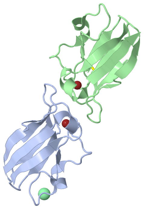
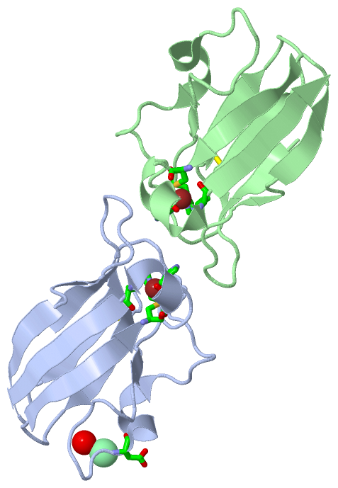
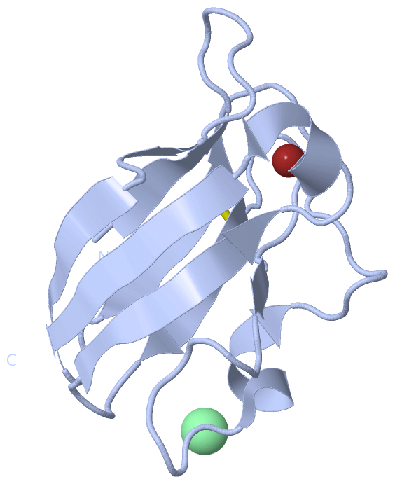
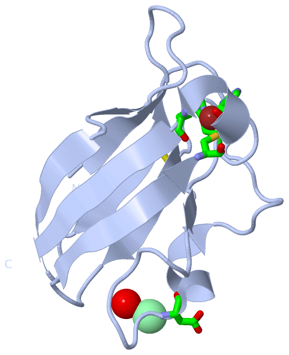
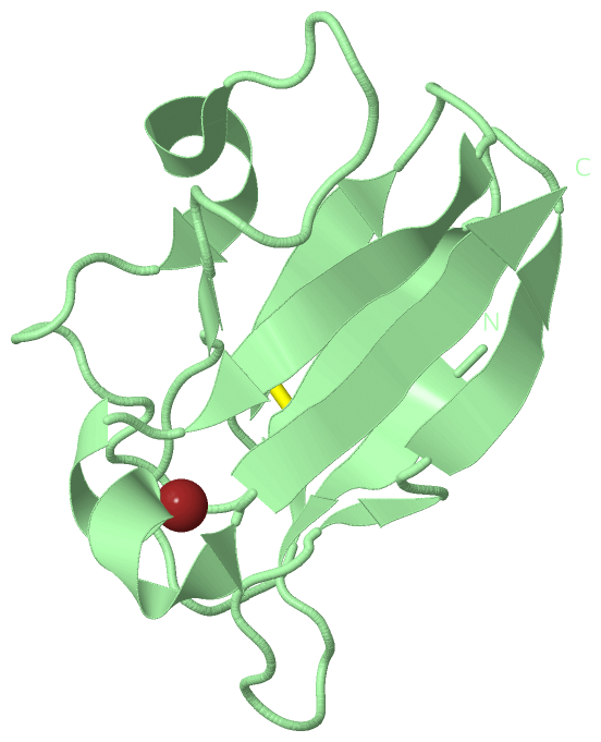
| | GO:0005507 | | copper ion binding | | Interacting selectively and non-covalently with copper (Cu) ions. |
| | GO:0009055 | | electron carrier activity | | Any molecular entity that serves as an electron acceptor and electron donor in an electron transport chain. An electron transport chain is a process in which a series of electron carriers operate together to transfer electrons from donors to any of several different terminal electron acceptors to generate a transmembrane electrochemical gradient. |
| | GO:0046872 | | metal ion binding | | Interacting selectively and non-covalently with any metal ion. |
| biological process |
|---|
| | GO:0055114 | | oxidation-reduction process | | A metabolic process that results in the removal or addition of one or more electrons to or from a substance, with or without the concomitant removal or addition of a proton or protons. |
| cellular component |
|---|
| | GO:0009507 | | chloroplast | | A chlorophyll-containing plastid with thylakoids organized into grana and frets, or stroma thylakoids, and embedded in a stroma. |
| | GO:0009535 | | chloroplast thylakoid membrane | | The pigmented membrane of a chloroplast thylakoid. An example of this component is found in Arabidopsis thaliana. |
| | GO:0016020 | | membrane | | A lipid bilayer along with all the proteins and protein complexes embedded in it an attached to it. |
| | GO:0009536 | | plastid | | Any member of a family of organelles found in the cytoplasm of plants and some protists, which are membrane-bounded and contain DNA. Plant plastids develop from a common type, the proplastid. |
| | GO:0009579 | | thylakoid | | A membranous cellular structure that bears the photosynthetic pigments in plants, algae, and cyanobacteria. In cyanobacteria thylakoids are of various shapes and are attached to, or continuous with, the plasma membrane. In eukaryotes they are flattened, membrane-bounded disk-like structures located in the chloroplasts; in the chloroplasts of higher plants the thylakoids form dense stacks called grana. Isolated thylakoid preparations can carry out photosynthetic electron transport and the associated phosphorylation. |
 Description