| molecular function |
|---|
| | GO:0020037 | | heme binding | | Interacting selectively and non-covalently with heme, any compound of iron complexed in a porphyrin (tetrapyrrole) ring. |
| | GO:0046872 | | metal ion binding | | Interacting selectively and non-covalently with any metal ion. |
| | GO:0005515 | | protein binding | | Interacting selectively and non-covalently with any protein or protein complex (a complex of two or more proteins that may include other nonprotein molecules). |
| biological process |
|---|
| | GO:0018063 | | cytochrome c-heme linkage | | The linkage of cytochromes and other heme proteins to heme. |
| | GO:0017004 | | cytochrome complex assembly | | The aggregation, arrangement and bonding together of a cytochrome complex. A cytochrome complex is a protein complex in which at least one of the proteins is a cytochrome, i.e. a heme-containing protein involved in catalysis of redox reactions. |
| | GO:0015886 | | heme transport | | The directed movement of heme, any compound of iron complexed in a porphyrin (tetrapyrrole) ring, into, out of or within a cell, or between cells, by means of some agent such as a transporter or pore. |
| | GO:0017003 | | protein-heme linkage | | The covalent linkage of heme and a protein. |
| cellular component |
|---|
| | GO:0016021 | | integral component of membrane | | The component of a membrane consisting of the gene products and protein complexes having at least some part of their peptide sequence embedded in the hydrophobic region of the membrane. |
| | GO:0031237 | | intrinsic component of periplasmic side of plasma membrane | | The component of a plasma membrane consisting of gene products and protein complexes that penetrate the periplasmic side of the plasma membrane only, either directly or via some covalently attached hydrophobic anchor. |
| | GO:0016020 | | membrane | | A lipid bilayer along with all the proteins and protein complexes embedded in it an attached to it. |
| | GO:0005886 | | plasma membrane | | The membrane surrounding a cell that separates the cell from its external environment. It consists of a phospholipid bilayer and associated proteins. |
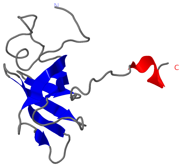
 Description
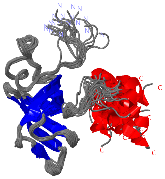