|
|
|
|
 Description
|
|
Compounds
|
||||||||||||||||||||
Chains, Units
Summary Information (see also Sequences/Alignments below) |
Ligands, Modified Residues, Ions (2, 8)| Asymmetric/Biological Unit (2, 8) |
Sites (8, 8)
Asymmetric Unit (8, 8)
|
SS Bonds (0, 0)| (no "SS Bond" information available for 1SAT) |
Cis Peptide Bonds (0, 0)| (no "Cis Peptide Bond" information available for 1SAT) |
SAPs(SNPs)/Variants (0, 0)| (no "SAP(SNP)/Variant" information available for 1SAT) |
PROSITE Motifs (2, 2)
Asymmetric/Biological Unit (2, 2)
|
||||||||||||||||||||||||||||||||
Exons (0, 0)| (no "Exon" information available for 1SAT) |
Sequences/Alignments
Asymmetric/Biological UnitChain A from PDB Type:PROTEIN Length:468 aligned with PRZN_SERMA | P23694 from UniProtKB/Swiss-Prot Length:487 Alignment length:468 29 39 49 59 69 79 89 99 109 119 129 139 149 159 169 179 189 199 209 219 229 239 249 259 269 279 289 299 309 319 329 339 349 359 369 379 389 399 409 419 429 439 449 459 469 479 PRZN_SERMA 20 TGYDAVDDLLHYHERGNGIQINGKDSFSNEQAGLFITRENQTWNGYKVFGQPVKLTFSFPDYKFSSTNVAGDTGLSKFSAEQQQQAKLSLQSWADVANITFTEVAAGQKANITFGNYSQDRPGHYDYGTQAYAFLPNTIWQGQDLGGQTWYNVNQSNVKHPATEDYGRQTFTHEIGHALGLSHPGDYNAGEGNPTYNDVTYAEDTRQFSLMSYWSETNTGGDNGGHYAAAPLLDDIAAIQHLYGANPSTRTGDTVYGFNSNTGRDFLSTTSNSQKVIFAAWDAGGNDTFDFSGYTANQRINLNEKSFSDVGGLKGNVSIAAGVTIENAIGGSGNDVIVGNAANNVLKGGAGNDVLFGGGGADELWGGAGKDIFVFSAASDSAPGASDWIRDFQKGIDKIDLSFFNKEANSSDFIHFVDHFSGTAGEALLSYNASSNVTDLSVNIGGHQAPDFLVKIVGQVDVATDFIV 487 SCOP domains d1sata2 A:4-246 Metalloprotease d1sata1 A:247-471 Metalloprotease SCOP domains CATH domains 1satA01 1satA02 A:18-249 Collagenase (Catalytic Domain) 1satA01 A:4-17,A:250-471 Alkaline Protease, subunit P, domain 1 CATH domains Pfam domains (1) ---------------------------------------------------------------Peptidase_M10-1satA01 A:67-209 -----------------------------------------------------------------------------------------------------------------------------------------------------HemolysinCabind-1s----------------------------------------------------------------------------------------------- Pfam domains (1) Pfam domains (2) -------------------------------------------------------------------------------------------------------------------------------------------------------------------------------------------------------------------------------------------------------------------------------------------------------------------------------------------------------------------HemolysinCabind-1s----------------------------------------------------------------------------------------------- Pfam domains (2) Pfam domains (3) -------------------------------------------------------------------------------------------------------------------------------------------------------------------------------------------------------------------------------------------------------------------------------------------------------------------------------------------------------------------HemolysinCabind-1s----------------------------------------------------------------------------------------------- Pfam domains (3) Pfam domains (4) -------------------------------------------------------------------------------------------------------------------------------------------------------------------------------------------------------------------------------------------------------------------------------------------------------------------------------------------------------------------HemolysinCabind-1s----------------------------------------------------------------------------------------------- Pfam domains (4) Pfam domains (5) ----------------------------------------------------------------------------------------------------------------------------------------------------------------------------------------------------------------------------------------------------Peptidase_M10_C-1satA06 A:248-471 Pfam domains (5) SAPs(SNPs) ------------------------------------------------------------------------------------------------------------------------------------------------------------------------------------------------------------------------------------------------------------------------------------------------------------------------------------------------------------------------------------------------------------------------------------------------------------------------------------ SAPs(SNPs) PROSITE -------------------------------------------------------------------------------------------------------------------------------------------------------------------------ZINC_PROTE-----------------------------------------------------------------------------------------------------------------------------------------------------------------------------HEMOLYSIN_CALCIUM ------------------------------------------------------------------------------------------------- PROSITE Transcript ------------------------------------------------------------------------------------------------------------------------------------------------------------------------------------------------------------------------------------------------------------------------------------------------------------------------------------------------------------------------------------------------------------------------------------------------------------------------------------ Transcript 1sat A 4 TGYDAVDDLLHYHERGNGIQINGKDSFSNEQAGLFITRENQTWNGYKVFGQPVKLTFSFPDYKFSSTNVAGDTGLSKFSAEQQQQAKLSLQSWADVANITFTEVAAGQKANITFGNYSQDRPGHYDYGTQAYAFLPNTIWQGQDLGGQTWYNVNQSNVKHPATEDYGRQTFTHEIGHALGLSHPGDYNAGEGDPTYADVTYAEDTRQFSLMSYWSETNTGGDNGGHYAAAPLLDDIAAIQHLYGANLSTRTGDTVYGFNSNTGRDFLSTTSNSQKVIFAAWDAGGNDTFDFSGYTANQRINLNEKSFSDVGGLKGNVSIAAGVTIENAIGGSGNDVIVGNAANNVLKGGAGNDVLFGGGGADELWGGAGKDIFVFSAASDSAPGASDWIRDFQKGIDKIDLSFFDKEANSSSFIHFVDHFSGTAGEALLSYNASSNVTDLSVNIGGHAAPDFLVKIVGQVDVATDFIV 471 13 23 33 43 53 63 73 83 93 103 113 123 133 143 153 163 173 183 193 203 213 223 233 243 253 263 273 283 293 303 313 323 333 343 353 363 373 383 393 403 413 423 433 443 453 463
|
||||||||||||||||||||
SCOP Domains (2, 2)
Asymmetric/Biological Unit

|
CATH Domains (2, 2)
Asymmetric/Biological Unit
|
Pfam Domains (3, 6)
Asymmetric/Biological Unit
|
Gene Ontology (12, 12)|
Asymmetric/Biological Unit(hide GO term definitions) Chain A (PRZN_SERMA | P23694)
|
||||||||||||||||||||||||||||||||||||||||||||||||||||||||||||||||||||||||||||||||||||||||||
Interactive Views
|
||||||||||||||||||||||||||||||||||||||||||||||||||||||||||||||||||||||||||||||||||||||||||||||||||||||||||||||||||||||||||||||||||||||||||||||||||||||||||||||||||||||||||||||
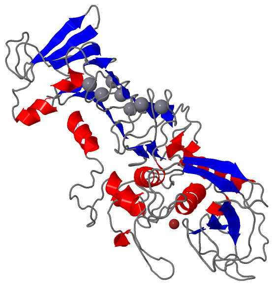
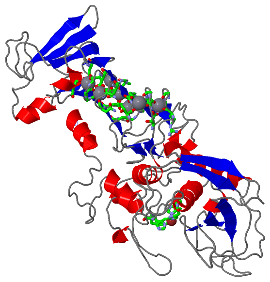
Still Images
|
||||||||||||||||
Databases
|
||||||||||||||||||||||||||||||||||||||||||||||||||||||||||||||||||||||||||||||||||||||||||||||||||||||||||||||||||||||||||||||||||||||||||||||||||||||||||||||||
Analysis Tools
|
|||||||||||||||||||||||||||||||||||||||||||||||||||||||||||||
Entries Sharing at Least One Protein Chain (UniProt ID)
Related Entries Specified in the PDB File
|
|