Show PDB file:   
         Plain Text   HTML   (compressed file size)
QuickSearch:   
by PDB,NDB,UniProt,PROSITE Code or Search Term(s)  
(-)Biol.Unit 1 - manually
(-)Asymmetric Unit
(-)Asym. Unit - sites
(-)Biological Unit 1
(-)Biol. Unit 1 - sites
(-)Biological Unit 2
(-)Biological Unit 3
collapse expand < >
Image Biol.Unit 1 - manually
Biol.Unit 1 - manually  (Jmol Viewer)
Image Asymmetric Unit
Asymmetric Unit  (Jmol Viewer)
Image Asym. Unit - sites
Asym. Unit - sites  (Jmol Viewer)
Image Biological Unit 1
Biological Unit 1  (Jmol Viewer)
Image Biol. Unit 1 - sites
Biol. Unit 1 - sites  (Jmol Viewer)
Image Biological Unit 2
Biological Unit 2  (Jmol Viewer)
Image Biological Unit 3
Biological Unit 3  (Jmol Viewer)

(-) Description

Title :  STRUCTURE OF OCR FROM BACTERIOPHAGE T7
 
Authors :  M. D. Walkinshaw, P. Taylor, S. S. Sturrock, C. Atanasiu, T. Berg, R. M. H J. M. Edwardson, D. T. Dryden
Date :  30 Jan 04  (Deposition) - 10 Feb 04  (Release) - 13 Jul 11  (Revision)
Method :  X-RAY DIFFRACTION
Resolution :  1.83
Chains :  Asym. Unit :  A
Biol. Unit 1:  A  (2x)
Biol. Unit 2:  A  (2x)
Biol. Unit 3:  A  (1x)
Keywords :  All Helical, Gene Regulation (Keyword Search: [Gene Ontology, PubMed, Web (Google))
 
Reference :  M. D. Walkinshaw, P. Taylor, S. S. Sturrock, C. Atanasiu, T. Berg, R. M. Henderson, J. M. Edwardson, D. T. Dryden
Structure Of Ocr From Bacteriophage T7, A Protein That Mimics B-Form Dna
Mol. Cell V. 9 187 2002
PubMed-ID: 11804597  |  Reference-DOI: 10.1016/S1097-2765(02)00435-5

(-) Compounds

Molecule 1 - GENE 0.3 PROTEIN
    ChainsA
    EngineeredYES
    Expression SystemESCHERICHIA COLI
    Expression System Taxid562
    Gene0.3
    Organism ScientificENTEROBACTERIA PHAGE T7
    Organism Taxid10760

 Structural Features

(-) Chains, Units

  1
Asymmetric Unit A
Biological Unit 1 (2x)A
Biological Unit 2 (2x)A
Biological Unit 3 (1x)A

Summary Information (see also Sequences/Alignments below)
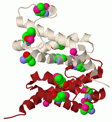
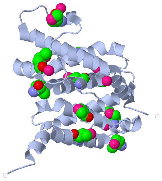
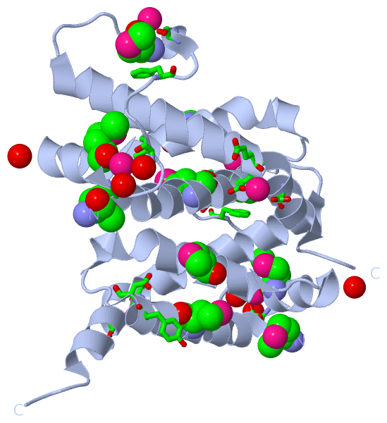
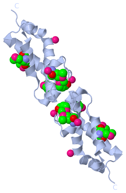
(-) Ligands, Modified Residues, Ions  (2, 8)

Asymmetric Unit (2, 8)
No.NameCountTypeFull Name
1CS3Ligand/IonCESIUM ION
2MSE5Mod. Amino AcidSELENOMETHIONINE
Biological Unit 1 (1, 10)
No.NameCountTypeFull Name
1CS-1Ligand/IonCESIUM ION
2MSE10Mod. Amino AcidSELENOMETHIONINE
Biological Unit 2 (1, 10)
No.NameCountTypeFull Name
1CS-1Ligand/IonCESIUM ION
2MSE10Mod. Amino AcidSELENOMETHIONINE
Biological Unit 3 (1, 5)
No.NameCountTypeFull Name
1CS-1Ligand/IonCESIUM ION
2MSE5Mod. Amino AcidSELENOMETHIONINE

(-) Sites  (3, 3)

Asymmetric Unit (3, 3)
No.NameEvidenceResiduesDescription
1AC1SOFTWARETYR A:24 , ASP A:25 , GLY A:69 , MSE A:71BINDING SITE FOR RESIDUE CS A 201
2AC2SOFTWAREMSE A:56 , SER A:58 , ILE A:61 , HOH A:253 , HOH A:255 , HOH A:309BINDING SITE FOR RESIDUE CS A 202
3AC3SOFTWAREPHE A:65 , SER A:68 , GLU A:95 , ASP A:96 , ASP A:99 , HOH A:285BINDING SITE FOR RESIDUE CS A 203

(-) SS Bonds  (0, 0)

(no "SS Bond" information available for 1S7Z)

(-) Cis Peptide Bonds  (0, 0)

(no "Cis Peptide Bond" information available for 1S7Z)

 Sequence-Structure Mapping

(-) SAPs(SNPs)/Variants  (0, 0)

(no "SAP(SNP)/Variant" information available for 1S7Z)

(-) PROSITE Motifs  (0, 0)

(no "PROSITE Motif" information available for 1S7Z)

(-) Exons   (0, 0)

(no "Exon" information available for 1S7Z)

(-) Sequences/Alignments

Asymmetric Unit
   Reformat: Number of residues per line =  ('0' or empty: single-line sequence representation)
  Number of residues per labelling interval =   
  UniProt sequence: complete  aligned part    
   Show mapping: SCOP domains CATH domains Pfam domains Secondary structure (by author)
SAPs(SNPs) PROSITE motifs Exons
(details for a mapped element are shown in a popup box when the mouse pointer rests over it)
Chain A from PDB  Type:PROTEIN  Length:106
 aligned with OCR_BPT7 | P03775 from UniProtKB/Swiss-Prot  Length:117

    Alignment length:106
                                    15        25        35        45        55        65        75        85        95       105      
             OCR_BPT7     6 MTYNNVFDHAYEMLKENIRYDDIRDTDDLHDAIHMAADNAVPHYYADIFSVMASEGIDLEFEDSGLMPDTKDVIRILQARIYEQLTIDLWEDAEDLLNEYLEEVEE 111
               SCOP domains d1s7za_ A: B-form DNA mimic Ocr                                                                            SCOP domains
               CATH domains ---------------------------------------------------------------------------------------------------------- CATH domains
               Pfam domains ---------------------------------------------------------------------------------------------------------- Pfam domains
         Sec.struct. author .hhhhhhhhhhhhhhhhhhhh...hhhhhhhhhhhhhhhhh..hhhhhhhhhh.........hhhhh....hhhhhhhhhhhhhhhhhhhhhhhhhhhhhhh.... Sec.struct. author
                 SAPs(SNPs) ---------------------------------------------------------------------------------------------------------- SAPs(SNPs)
                    PROSITE ---------------------------------------------------------------------------------------------------------- PROSITE
                 Transcript ---------------------------------------------------------------------------------------------------------- Transcript
                 1s7z A   5 mTYNNVFDHAYEmLKENIRYDDIRDTDDLHDAIHmAADNAVPHYYADIFSVmASEGIDLEFEDSGLmPDTKDVIRILQARIYEQLTIDLWEDAEDLLNEYLEEVEE 110
                            |       14  |     24        34    |   44        54 |      64      | 74        84        94       104      
                            |          17-MSE                39-MSE           56-MSE         71-MSE                                   
                            5-MSE                                                                                                     
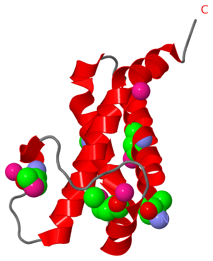
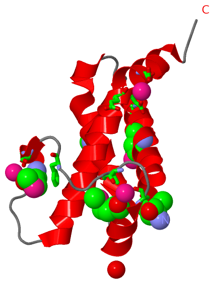
   Legend:   → Mismatch (orange background)
  - → Gap (green background, '-', border residues have a numbering label)
    → Modified Residue (blue background, lower-case, 'x' indicates undefined single-letter code, labelled with number + name)
  x → Chemical Group (purple background, 'x', labelled with number + name, e.g. ACE or NH2)
  extra numbering lines below/above indicate numbering irregularities and modified residue names etc., number ends below/above '|'

 Classification and Annotation

(-) SCOP Domains  (1, 1)

Asymmetric Unit

(-) CATH Domains  (0, 0)

(no "CATH Domain" information available for 1S7Z)

(-) Pfam Domains  (0, 0)

(no "Pfam Domain" information available for 1S7Z)

(-) Gene Ontology  (1, 1)

Asymmetric Unit(hide GO term definitions)
Chain A   (OCR_BPT7 | P03775)
biological process
    GO:0016032    viral process    A multi-organism process in which a virus is a participant. The other participant is the host. Includes infection of a host cell, replication of the viral genome, and assembly of progeny virus particles. In some cases the viral genetic material may integrate into the host genome and only subsequently, under particular circumstances, 'complete' its life cycle.

 Visualization

(-) Interactive Views

Asymmetric Unit
  Complete Structure
    Jena3D(integrated viewing of ligand, site, SAP, PROSITE, SCOP information)
    WebMol | AstexViewer[tm]@PDBe
(Java Applets, require no local installation except for Java; loading may be slow)
    STRAP
(Java WebStart application, automatic local installation, requires Java; full application with system access!)
    RasMol
(require local installation)
    Molscript (VRML)
(requires installation of a VRML viewer; select preferred view via VRML and generate a mono or stereo PDF format file)
 
  Ligands, Modified Residues, Ions
    CS  [ RasMol | Jena3D ]  +environment [ RasMol | Jena3D ]
    MSE  [ RasMol | Jena3D ]  +environment [ RasMol | Jena3D ]
 
  Sites
    AC1  [ RasMol ]  +environment [ RasMol ]
    AC2  [ RasMol ]  +environment [ RasMol ]
    AC3  [ RasMol ]  +environment [ RasMol ]
 
  Cis Peptide Bonds
(no "Cis Peptide Bonds" information available for 1s7z)
 
Biological Units
  Complete Structure
    Biological Unit 1  [ Jena3D ]
    Biological Unit 2  [ Jena3D ]
    Biological Unit 3  [ Jena3D ]

(-) Still Images

Jmol
  protein: cartoon or spacefill or dots and stick; nucleic acid: cartoon and stick; ligands: spacefill; active site: stick
Molscript
  protein, nucleic acid: cartoon; ligands: spacefill; active site: ball and stick

 Databases and Analysis Tools

(-) Databases

Access by PDB/NDB ID
  1s7z
    Family and Domain InformationProDom | SYSTERS
    General Structural InformationGlycoscienceDB | MMDB | NDB | OCA | PDB | PDBe | PDBj | PDBsum | PDBWiki | PQS | PROTEOPEDIA
    Orientation in MembranesOPM
    Protein SurfaceSURFACE
    Secondary StructureDSSP (structure derived) | HSSP (homology derived)
    Structural GenomicsGeneCensus
    Structural NeighboursCE | VAST
    Structure ClassificationCATH | Dali | SCOP
    Validation and Original DataBMRB Data View | BMRB Restraints Grid | EDS | PROCHECK | RECOORD | WHAT_CHECK
 
Access by UniProt ID/Accession number
  OCR_BPT7 | P03775
    Comparative Protein Structure ModelsModBase
    Genomic InformationEnsembl
    Protein-protein InteractionDIP
    Sequence, Family and Domain InformationInterPro | Pfam | SMART | UniProtKB/SwissProt
 
Access by Enzyme Classificator   (EC Number)
  (no 'Enzyme Classificator' available)
    General Enzyme InformationBRENDA | EC-PDB | Enzyme | IntEnz
    PathwayKEGG | MetaCyc
 
Access by Disease Identifier   (MIM ID)
  (no 'MIM ID' available)
    Disease InformationOMIM
 
Access by GenAge ID
  (no 'GenAge ID' available)
    Age Related InformationGenAge

(-) Analysis Tools

Access by PDB/NDB ID
    Domain InformationXDom
    Interatomic Contacts of Structural UnitsCSU
    Ligand-protein ContactsLPC
    Protein CavitiescastP
    Sequence and Secondary StructurePDBCartoon
    Structure AlignmentSTRAP(Java WebStart application, automatic local installation, requires Java; full application with system access!)
    Structure and Sequence BrowserSTING
 
Access by UniProt ID/Accession number
  OCR_BPT7 | P03775
    Protein Disorder PredictionDisEMBL | FoldIndex | GLOBPLOT (for more information see DisProt)

 Related Entries

(-) Entries Sharing at Least One Protein Chain (UniProt ID)

UniProtKB/Swiss-Prot
        OCR_BPT7 | P037752y7c

(-) Related Entries Specified in the PDB File

(no "Related Entries Specified in the PDB File" available for 1S7Z)