|
|
|
|
 Description
|
|
Compounds
|
||||||||||||||||||||||||||||||||||||||||||||
Chains, Units
Summary Information (see also Sequences/Alignments below) |
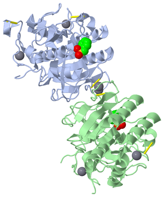
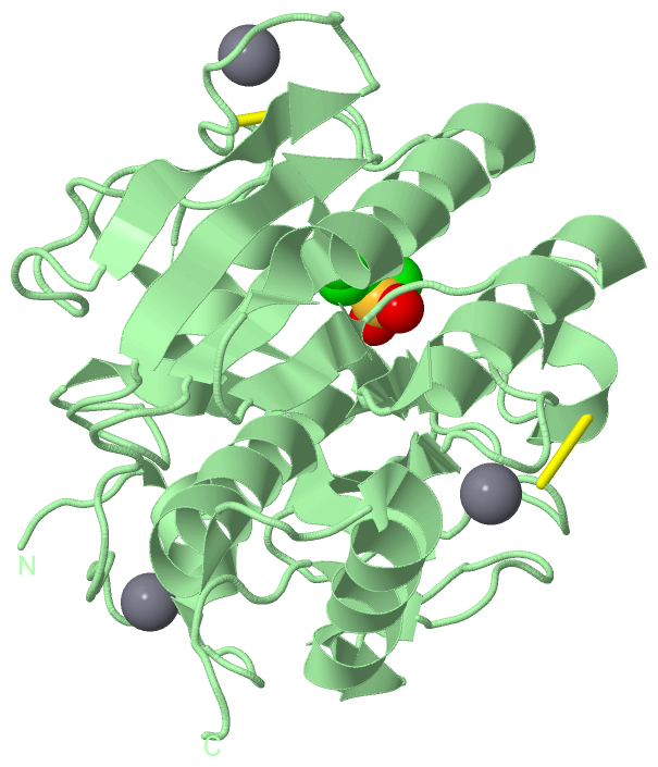
Ligands, Modified Residues, Ions (2, 8)| Asymmetric Unit (2, 8) Biological Unit 1 (1, 1) Biological Unit 2 (1, 1) |
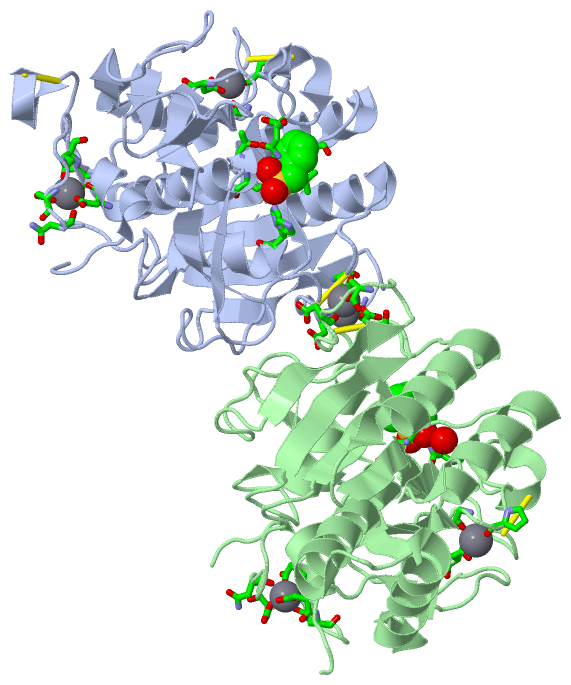
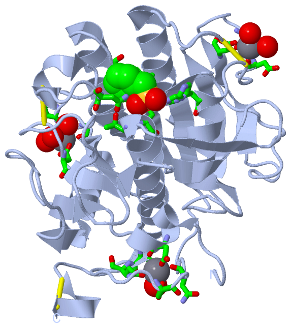
Sites (8, 8)
Asymmetric Unit (8, 8)
|
SS Bonds (5, 5)
Asymmetric Unit
|
||||||||||||||||||||||||
Cis Peptide Bonds (4, 4)
Asymmetric Unit
|
||||||||||||||||||||
SAPs(SNPs)/Variants (0, 0)| (no "SAP(SNP)/Variant" information available for 1S2N) |
PROSITE Motifs (0, 0)| (no "PROSITE Motif" information available for 1S2N) |
Exons (0, 0)| (no "Exon" information available for 1S2N) |
Sequences/Alignments
Asymmetric UnitChain A from PDB Type:PROTEIN Length:281 aligned with Q8GB52_9VIBR | Q8GB52 from UniProtKB/TrEMBL Length:530 Alignment length:281 149 159 169 179 189 199 209 219 229 239 249 259 269 279 289 299 309 319 329 339 349 359 369 379 389 399 409 419 Q8GB52_9VIBR 140 QSNAIWGLDRIDQRNLPLDRNYNANFDGFGVTAYVIDTGVNNNHEEFGGRSVSGYDFVDNDADSSDCNGHGTHVAGTIGGSQYGVAKNVNIVGVRVLSCSGSGTTSGVISGVDWVAQNASGPSVANMSLGGGQSTALDSAVQGAIQSGVSFMLAAGNSNADACNTSPARVPSGVTVGSTTSSDSRSSFSNWGSCVDLFAPGSQIKSAWYDGGYKTISGTSMATPHVAGVAALYLQENNGLTPLQLTGLLNSRASENKVSDTRGTTNKLLYSLADSGCEPDC 420 SCOP domains d1s2na_ A: automated matches SCOP domains CATH domains 1s2nA00 A:1-281 [code=3.40.50.200, no name defined] CATH domains Pfam domains ----------------------------------------------------------------------------------------------------------------------------------------------------------------------------------------------------------------------------------------------------------------------------------------- Pfam domains SAPs(SNPs) ----------------------------------------------------------------------------------------------------------------------------------------------------------------------------------------------------------------------------------------------------------------------------------------- SAPs(SNPs) PROSITE ----------------------------------------------------------------------------------------------------------------------------------------------------------------------------------------------------------------------------------------------------------------------------------------- PROSITE Transcript ----------------------------------------------------------------------------------------------------------------------------------------------------------------------------------------------------------------------------------------------------------------------------------------- Transcript 1s2n A 1 QSNAIWGLDRIDQRNLPLDRNYNANFDGFGVTAYVIDTGVNNNHEEFGGRSVSGYDFVDNDADSSDCNGHGTHVAGTIGGSQYGVAKNVNIVGVRVLSCSGSGTTSGVISGVDWVAQNASGPSVANMSLGGGQSTALDSAVQGAIQSGVSFMLAAGNSNADACNTSPARVPSGVTVGSTTSSDSRSSFSNWGSCVDLFAPGSQIKSAWYDGGYKTISGTSMATPHVAGVAALYLQENNGLTPLQLTGLLNSRASENKVSDTRGTTNKLLYSLADSGCEPDC 281 10 20 30 40 50 60 70 80 90 100 110 120 130 140 150 160 170 180 190 200 210 220 230 240 250 260 270 280 Chain B from PDB Type:PROTEIN Length:273 aligned with Q8GB52_9VIBR | Q8GB52 from UniProtKB/TrEMBL Length:530 Alignment length:273 150 160 170 180 190 200 210 220 230 240 250 260 270 280 290 300 310 320 330 340 350 360 370 380 390 400 410 Q8GB52_9VIBR 141 SNAIWGLDRIDQRNLPLDRNYNANFDGFGVTAYVIDTGVNNNHEEFGGRSVSGYDFVDNDADSSDCNGHGTHVAGTIGGSQYGVAKNVNIVGVRVLSCSGSGTTSGVISGVDWVAQNASGPSVANMSLGGGQSTALDSAVQGAIQSGVSFMLAAGNSNADACNTSPARVPSGVTVGSTTSSDSRSSFSNWGSCVDLFAPGSQIKSAWYDGGYKTISGTSMATPHVAGVAALYLQENNGLTPLQLTGLLNSRASENKVSDTRGTTNKLLYSLAD 413 SCOP domains d1s2nb_ B: automated matches SCOP domains CATH domains 1s2nB00 B:2-274 [code=3.40.50.200, no name defined] CATH domains Pfam domains (1) ------------------------------Peptidase_S8-1s2nB01 B:32-274 Pfam domains (1) Pfam domains (2) ------------------------------Peptidase_S8-1s2nB02 B:32-274 Pfam domains (2) SAPs(SNPs) --------------------------------------------------------------------------------------------------------------------------------------------------------------------------------------------------------------------------------------------------------------------------------- SAPs(SNPs) PROSITE --------------------------------------------------------------------------------------------------------------------------------------------------------------------------------------------------------------------------------------------------------------------------------- PROSITE Transcript --------------------------------------------------------------------------------------------------------------------------------------------------------------------------------------------------------------------------------------------------------------------------------- Transcript 1s2n B 2 SNAIWGLDRIDQRNLPLDRNYNANFDGFGVTAYVIDTGVNNNHEEFGGRSVSGYDFVDNDADSSDCNGHGTHVAGTIGGSQYGVAKNVNIVGVRVLSCSGSGTTSGVISGVDWVAQNASGPSVANMSLGGGQSTALDSAVQGAIQSGVSFMLAAGNSNADACNTSPARVPSGVTVGSTTSSDSRSSFSNWGSCVDLFAPGSQIKSAWYDGGYKTISGTSMATPHVAGVAALYLQENNGLTPLQLTGLLNSRASENKVSDTRGTTNKLLYSLAD 274 11 21 31 41 51 61 71 81 91 101 111 121 131 141 151 161 171 181 191 201 211 221 231 241 251 261 271
|
||||||||||||||||||||
SCOP Domains (1, 2)
Asymmetric Unit
 |
CATH Domains (1, 2)
Asymmetric Unit
|
Pfam Domains (1, 2)
Asymmetric Unit
|
Gene Ontology (6, 6)|
Asymmetric Unit(hide GO term definitions) Chain A,B (Q8GB52_9VIBR | Q8GB52)
|
||||||||||||||||||||||||||||||||||||||||||||||||
Interactive Views
|
|||||||||||||||||||||||||||||||||||||||||||||||||||||||||||||||||||||||||||||||||||||||||||||||||||||||||||||||||||||||||||||||||||||||||||||||||||||||||||||||||||||||||||||||||||||||||||||||||||||||||||||||||||||||||||
Still Images
|
||||||||||||||||
Databases
|
||||||||||||||||||||||||||||||||||||||||||||||||||||||||||||||||||||||||||||||||||||||||||||||||||||||||||||||||||||||||||||||||||||||||||||||||||||||||||||||||
Analysis Tools
|
|||||||||||||||||||||||||||||||||||||||||||||||||||||||||||||
Entries Sharing at Least One Protein Chain (UniProt ID)
Related Entries Specified in the PDB File
|
|