|
|
|
|
 Description
|
|
Compounds
|
||||||||||||||||||||||||||||||||||||||||||||||||||||
Chains, Units
Summary Information (see also Sequences/Alignments below) |
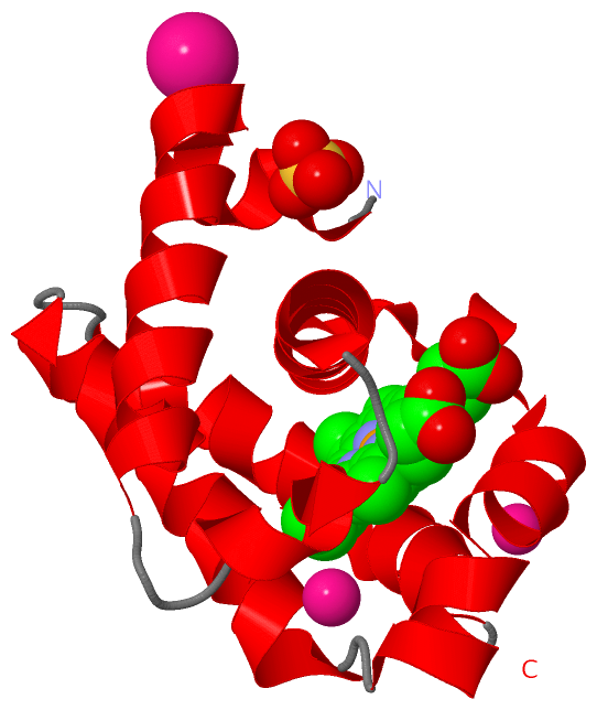
Ligands, Modified Residues, Ions (4, 5)| Asymmetric/Biological Unit (4, 5) |
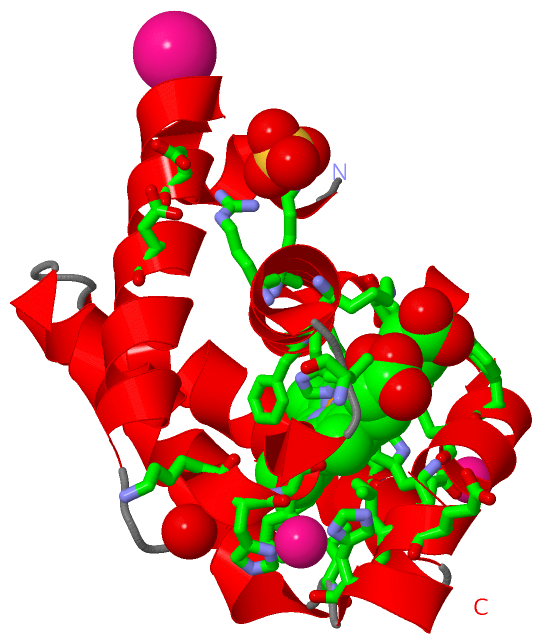
Sites (5, 5)
Asymmetric Unit (5, 5)
|
SS Bonds (0, 0)| (no "SS Bond" information available for 1RTX) |
Cis Peptide Bonds (0, 0)| (no "Cis Peptide Bond" information available for 1RTX) |
SAPs(SNPs)/Variants (0, 0)| (no "SAP(SNP)/Variant" information available for 1RTX) |
PROSITE Motifs (1, 1)
Asymmetric/Biological Unit (1, 1)
|
||||||||||||||||||||||||
Exons (0, 0)| (no "Exon" information available for 1RTX) |
Sequences/Alignments
Asymmetric/Biological UnitChain A from PDB Type:PROTEIN Length:123 aligned with TRHBN_SYNY3 | P73925 from UniProtKB/Swiss-Prot Length:124 Alignment length:123 11 21 31 41 51 61 71 81 91 101 111 121 TRHBN_SYNY3 2 STLYEKLGGTTAVDLAVDKFYERVLQDDRIKHFFADVDMAKQRAHQKAFLTYAFGGTDKYDGRYMREAHKELVENHGLNGEHFDAVAEDLLATLKEMGVPEDLIAEVAAVAGAPAHKRDVLNQ 124 SCOP domains d1rtxa_ A: Protozoan/bacterial hemoglobin SCOP domains CATH domains 1rtxA00 A:2-124 Globins CATH domains Pfam domains -Bac_globin-1rtxA01 A:3-121 --- Pfam domains SAPs(SNPs) --------------------------------------------------------------------------------------------------------------------------- SAPs(SNPs) PROSITE ------------------------------------------------GLOBIN_FAM_2 ------------------------------------------------------ PROSITE Transcript --------------------------------------------------------------------------------------------------------------------------- Transcript 1rtx A 2 STLYEKLGGTTAVDLAVDKFYERVLQDDRIKHFFADVDMAKQRAHQKAFLTYAFGGTDKYDGRYMREAHKELVENHGLNGEHFDAVAEDLLATLKEMGVPEDLIAEVAAVAGAPAHKRDVLNQ 124 11 21 31 41 51 61 71 81 91 101 111 121
|
||||||||||||||||||||
SCOP Domains (1, 1)
Asymmetric/Biological Unit
 |
CATH Domains (1, 1)
Asymmetric/Biological Unit
|
Pfam Domains (1, 1)
Asymmetric/Biological Unit
|
Gene Ontology (6, 6)|
Asymmetric/Biological Unit(hide GO term definitions) Chain A (TRHBN_SYNY3 | P73925)
|
||||||||||||||||||||||||||||||||||||||||||||||||
Interactive Views
|
|||||||||||||||||||||||||||||||||||||||||||||||||||||||||||||||||||||||||||||||||||||||||||||||||||||||||||||||||||||||||||||||||||||||||||||||||||||||||||||||||||||||
Still Images
|
||||||||||||||||
Databases
|
||||||||||||||||||||||||||||||||||||||||||||||||||||||||||||||||||||||||||||||||||||||||||||||||||||||||||||||||||||||||||||||||||||||||||||||||||||||||||||||||
Analysis Tools
|
|||||||||||||||||||||||||||||||||||||||||||||||||||||||||||||
Entries Sharing at Least One Protein Chain (UniProt ID)
Related Entries Specified in the PDB File
|
|