|
|
|
|
 Description
|
|
Compounds
|
||||||||||||||||||||||||
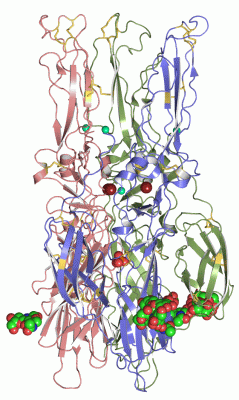
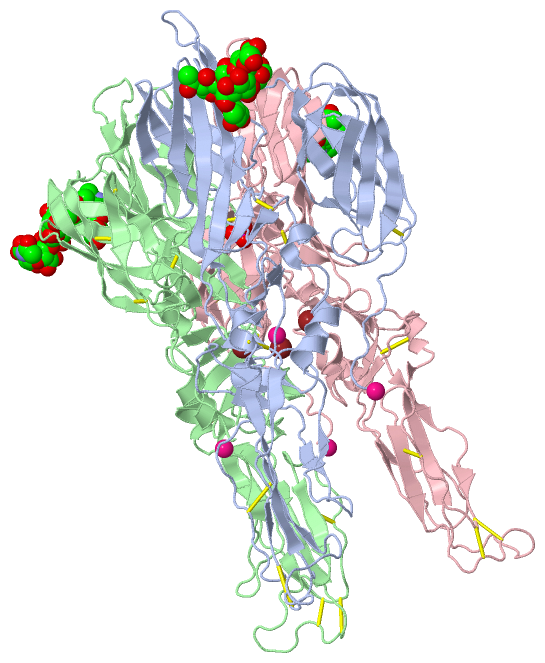
Chains, Units
Summary Information (see also Sequences/Alignments below) |
Ligands, Modified Residues, Ions (9, 23)
Asymmetric Unit (9, 23)
|
Sites (21, 21)
Asymmetric Unit (21, 21)
|
SS Bonds (24, 24)
Asymmetric Unit
|
||||||||||||||||||||||||||||||||||||||||||||||||||||||||||||||||||||||||||||||||||||||||||||||||||||
Cis Peptide Bonds (0, 0)| (no "Cis Peptide Bond" information available for 1RER) |
SAPs(SNPs)/Variants (8, 24)
Asymmetric Unit (8, 24)
|
|||||||||||||||||||||||||||||||||||||||||||||||||||||||||||||||||||||||||||||||||||||||||||||||||||||||||||||||||||||||||||||||||||||||||||||||||||||||||||||||||||||||||||||||||||||||||||||||||||||||||||||||||||||||||||||||||||||||||||||||||||||||||||||||||||||||||||||||||||||||||||||||||||||||||||||||||||||||||||||||||||||||||||||||||||||||||||||||||||||||||||||||||||||||||||||||||||||||||||||||||||||||||||||||||||||||||||||||||||||||||||||||
PROSITE Motifs (0, 0)| (no "PROSITE Motif" information available for 1RER) |
Exons (0, 0)| (no "Exon" information available for 1RER) |
Sequences/Alignments
Asymmetric UnitChain A from PDB Type:PROTEIN Length:391 aligned with POLS_SFV | P03315 from UniProtKB/Swiss-Prot Length:1253 Alignment length:391 825 835 845 855 865 875 885 895 905 915 925 935 945 955 965 975 985 995 1005 1015 1025 1035 1045 1055 1065 1075 1085 1095 1105 1115 1125 1135 1145 1155 1165 1175 1185 1195 1205 POLS_SFV 816 YEHSTVMPNVVGFPYKAHIERPGYSPLTLQMQVVETSLEPTLNLEYITCEYKTVVPSPYVKCCGASECSTKEKPDYQCKVYTGVYPFMWGGAYCFCDSENTQLSEAYVDRSDVCRHDHASAYKAHTASLKAKVRVMYGNVNQTVDVYVNGDHAVTIGGTQFIFGPLSSAWTPFDNKIVVYKDEVFNQDFPPYGSGQPGRFGDIQSRTVESNDLYANTALKLARPSPGMVHVPYTQTPSGFKYWLKEKGTALNTKAPFGCQIKTNPVRAMNCAVGNIPVSMNLPDSAFTRIVEAPTIIDLTCTVATCTHSSDFGGVLTLTYKTNKNGDCSVHSHSNVATLQEATAKVKTAGKVTLHFSTASASPSFVVSLCSARATCSASCEPPKDHIVPYA 1206 SCOP domains d1rera2 A:1-292 Fusion glycoprotein E1 d1rera1 A:293-391 Fusion glycoprotein E1 SCOP domains CATH domains 1rerA01 A:1-42,A:124-173,A:257-290 ---------------------------------------------------------------------------------1rerA01 A:1-42,A:124-173,A:257-290 -----------------------------------------------------------------------------------1rerA01 A:1-42,A:124-173,A:257-290----------------------------------------------------------------------------------------------------- CATH domains Pfam domains ------------------------------------------------------------------------------------------------------------------------------------------------------------------------------------------------------------------------------------------------------------------------------------------------------------------------------------------------------------------------------------------------------- Pfam domains SAPs(SNPs) ----------------------------------------------------------------S-------------------------------------------------K----------------------------------------------------------------------------------------------------------------T--------------------------------------------------------------------T---------------------K---D--------------------------R----------------------K------------------ SAPs(SNPs) PROSITE ------------------------------------------------------------------------------------------------------------------------------------------------------------------------------------------------------------------------------------------------------------------------------------------------------------------------------------------------------------------------------------------------------- PROSITE Transcript ------------------------------------------------------------------------------------------------------------------------------------------------------------------------------------------------------------------------------------------------------------------------------------------------------------------------------------------------------------------------------------------------------- Transcript 1rer A 1 YEHSTVMPNVVGFPYKAHIERPGYSPLTLQMQVVETSLEPTLNLEYITCEYKTVVPSPYVKCCGASECSTKEKPDYQCKVYTGVYPFMWGGAYCFCDSENTQLSEAYVDRSDVCRHDHASAYKAHTASLKAKVRVMYGNVNQTVDVYVNGDHAVTIGGTQFIFGPLSSAWTPFDNKIVVYKDEVFNQDFPPYGSGQPGRFGDIQSRTVESNDLYANTALKLARPSPGMVHVPYTQTPSGFKYWLKEKGTALNTKAPFGCQIKTNPVRAMNCAVGNIPVSMNLPDSAFTRIVEAPTIIDLTCTVATCTHSSDFGGVLTLTYKTNKNGDCSVHSHSNVATLQEATAKVKTAGKVTLHFSTASASPSFVVSLCSARATCSASCEPPKDHIVPYA 391 10 20 30 40 50 60 70 80 90 100 110 120 130 140 150 160 170 180 190 200 210 220 230 240 250 260 270 280 290 300 310 320 330 340 350 360 370 380 390 Chain B from PDB Type:PROTEIN Length:391 aligned with POLS_SFV | P03315 from UniProtKB/Swiss-Prot Length:1253 Alignment length:391 825 835 845 855 865 875 885 895 905 915 925 935 945 955 965 975 985 995 1005 1015 1025 1035 1045 1055 1065 1075 1085 1095 1105 1115 1125 1135 1145 1155 1165 1175 1185 1195 1205 POLS_SFV 816 YEHSTVMPNVVGFPYKAHIERPGYSPLTLQMQVVETSLEPTLNLEYITCEYKTVVPSPYVKCCGASECSTKEKPDYQCKVYTGVYPFMWGGAYCFCDSENTQLSEAYVDRSDVCRHDHASAYKAHTASLKAKVRVMYGNVNQTVDVYVNGDHAVTIGGTQFIFGPLSSAWTPFDNKIVVYKDEVFNQDFPPYGSGQPGRFGDIQSRTVESNDLYANTALKLARPSPGMVHVPYTQTPSGFKYWLKEKGTALNTKAPFGCQIKTNPVRAMNCAVGNIPVSMNLPDSAFTRIVEAPTIIDLTCTVATCTHSSDFGGVLTLTYKTNKNGDCSVHSHSNVATLQEATAKVKTAGKVTLHFSTASASPSFVVSLCSARATCSASCEPPKDHIVPYA 1206 SCOP domains d1rerb2 B:1-292 Fusion glycoprotein E1 d1rerb1 B:293-391 Fusion glycoprotein E1 SCOP domains CATH domains 1rerB01 B:1-42,B:124-173,B:257-290 ---------------------------------------------------------------------------------1rerB01 B:1-42,B:124-173,B:257-290 -----------------------------------------------------------------------------------1rerB01 B:1-42,B:124-173,B:257-290----------------------------------------------------------------------------------------------------- CATH domains Pfam domains ------------------------------------------------------------------------------------------------------------------------------------------------------------------------------------------------------------------------------------------------------------------------------------------------------------------------------------------------------------------------------------------------------- Pfam domains SAPs(SNPs) ----------------------------------------------------------------S-------------------------------------------------K----------------------------------------------------------------------------------------------------------------T--------------------------------------------------------------------T---------------------K---D--------------------------R----------------------K------------------ SAPs(SNPs) PROSITE ------------------------------------------------------------------------------------------------------------------------------------------------------------------------------------------------------------------------------------------------------------------------------------------------------------------------------------------------------------------------------------------------------- PROSITE Transcript ------------------------------------------------------------------------------------------------------------------------------------------------------------------------------------------------------------------------------------------------------------------------------------------------------------------------------------------------------------------------------------------------------- Transcript 1rer B 1 YEHSTVMPNVVGFPYKAHIERPGYSPLTLQMQVVETSLEPTLNLEYITCEYKTVVPSPYVKCCGASECSTKEKPDYQCKVYTGVYPFMWGGAYCFCDSENTQLSEAYVDRSDVCRHDHASAYKAHTASLKAKVRVMYGNVNQTVDVYVNGDHAVTIGGTQFIFGPLSSAWTPFDNKIVVYKDEVFNQDFPPYGSGQPGRFGDIQSRTVESNDLYANTALKLARPSPGMVHVPYTQTPSGFKYWLKEKGTALNTKAPFGCQIKTNPVRAMNCAVGNIPVSMNLPDSAFTRIVEAPTIIDLTCTVATCTHSSDFGGVLTLTYKTNKNGDCSVHSHSNVATLQEATAKVKTAGKVTLHFSTASASPSFVVSLCSARATCSASCEPPKDHIVPYA 391 10 20 30 40 50 60 70 80 90 100 110 120 130 140 150 160 170 180 190 200 210 220 230 240 250 260 270 280 290 300 310 320 330 340 350 360 370 380 390 Chain C from PDB Type:PROTEIN Length:391 aligned with POLS_SFV | P03315 from UniProtKB/Swiss-Prot Length:1253 Alignment length:391 825 835 845 855 865 875 885 895 905 915 925 935 945 955 965 975 985 995 1005 1015 1025 1035 1045 1055 1065 1075 1085 1095 1105 1115 1125 1135 1145 1155 1165 1175 1185 1195 1205 POLS_SFV 816 YEHSTVMPNVVGFPYKAHIERPGYSPLTLQMQVVETSLEPTLNLEYITCEYKTVVPSPYVKCCGASECSTKEKPDYQCKVYTGVYPFMWGGAYCFCDSENTQLSEAYVDRSDVCRHDHASAYKAHTASLKAKVRVMYGNVNQTVDVYVNGDHAVTIGGTQFIFGPLSSAWTPFDNKIVVYKDEVFNQDFPPYGSGQPGRFGDIQSRTVESNDLYANTALKLARPSPGMVHVPYTQTPSGFKYWLKEKGTALNTKAPFGCQIKTNPVRAMNCAVGNIPVSMNLPDSAFTRIVEAPTIIDLTCTVATCTHSSDFGGVLTLTYKTNKNGDCSVHSHSNVATLQEATAKVKTAGKVTLHFSTASASPSFVVSLCSARATCSASCEPPKDHIVPYA 1206 SCOP domains d1rerc2 C:1-292 Fusion glycoprotein E1 d1rerc1 C:293-391 Fusion glycoprotein E1 SCOP domains CATH domains 1rerC01 C:1-42,C:124-173,C:257-290 ---------------------------------------------------------------------------------1rerC01 C:1-42,C:124-173,C:257-290 -----------------------------------------------------------------------------------1rerC01 C:1-42,C:124-173,C:257-290----------------------------------------------------------------------------------------------------- CATH domains Pfam domains (1) Alpha_E1_glycop-1rerC01 C:1-391 Pfam domains (1) Pfam domains (2) Alpha_E1_glycop-1rerC02 C:1-391 Pfam domains (2) Pfam domains (3) Alpha_E1_glycop-1rerC03 C:1-391 Pfam domains (3) SAPs(SNPs) ----------------------------------------------------------------S-------------------------------------------------K----------------------------------------------------------------------------------------------------------------T--------------------------------------------------------------------T---------------------K---D--------------------------R----------------------K------------------ SAPs(SNPs) PROSITE ------------------------------------------------------------------------------------------------------------------------------------------------------------------------------------------------------------------------------------------------------------------------------------------------------------------------------------------------------------------------------------------------------- PROSITE Transcript ------------------------------------------------------------------------------------------------------------------------------------------------------------------------------------------------------------------------------------------------------------------------------------------------------------------------------------------------------------------------------------------------------- Transcript 1rer C 1 YEHSTVMPNVVGFPYKAHIERPGYSPLTLQMQVVETSLEPTLNLEYITCEYKTVVPSPYVKCCGASECSTKEKPDYQCKVYTGVYPFMWGGAYCFCDSENTQLSEAYVDRSDVCRHDHASAYKAHTASLKAKVRVMYGNVNQTVDVYVNGDHAVTIGGTQFIFGPLSSAWTPFDNKIVVYKDEVFNQDFPPYGSGQPGRFGDIQSRTVESNDLYANTALKLARPSPGMVHVPYTQTPSGFKYWLKEKGTALNTKAPFGCQIKTNPVRAMNCAVGNIPVSMNLPDSAFTRIVEAPTIIDLTCTVATCTHSSDFGGVLTLTYKTNKNGDCSVHSHSNVATLQEATAKVKTAGKVTLHFSTASASPSFVVSLCSARATCSASCEPPKDHIVPYA 391 10 20 30 40 50 60 70 80 90 100 110 120 130 140 150 160 170 180 190 200 210 220 230 240 250 260 270 280 290 300 310 320 330 340 350 360 370 380 390
|
||||||||||||||||||||
SCOP Domains (2, 6)
Asymmetric Unit
 |
CATH Domains (1, 3)
Asymmetric Unit
|
Pfam Domains (1, 3)
Asymmetric Unit
|
Gene Ontology (26, 26)|
Asymmetric Unit(hide GO term definitions) Chain A,B,C (POLS_SFV | P03315)
|
||||||||||||||||||||||||||||||||||||||||||||||||||||||||||||||||||||||||||||||||||||||||||||||||||||||||||||||||||||||||||||||||||||||||||||||||||||||||||||||||||||||||||||||
Interactive Views
|
|||||||||||||||||||||||||||||||||||||||||||||||||||||||||||||||||||||||||||||||||||||||||||||||||||||||||||||||||||||||||||||||||||||||||||||||||||||||||||||||||||||||||||||||||||||||||||||||||||||||||||||||||||||||||||||||||||||||||||||||||||||||||||||||||||||||||||||||||||||||||||||||||||||||||||||||||||||||||||||||||||||||||||||||||
Still Images
|
||||||||||||||||
Databases
|
||||||||||||||||||||||||||||||||||||||||||||||||||||||||||||||||||||||||||||||||||||||||||||||||||||||||||||||||||||||||||||||||||||||||||||||||||||||||||||||||
Analysis Tools
|
|||||||||||||||||||||||||||||||||||||||||||||||||||||||||||||
Entries Sharing at Least One Protein Chain (UniProt ID)
Related Entries Specified in the PDB File
|
|