|
|
|
|
 Description
|
|
Compounds
|
||||||||||||||||||||||||
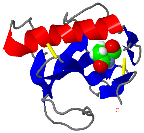
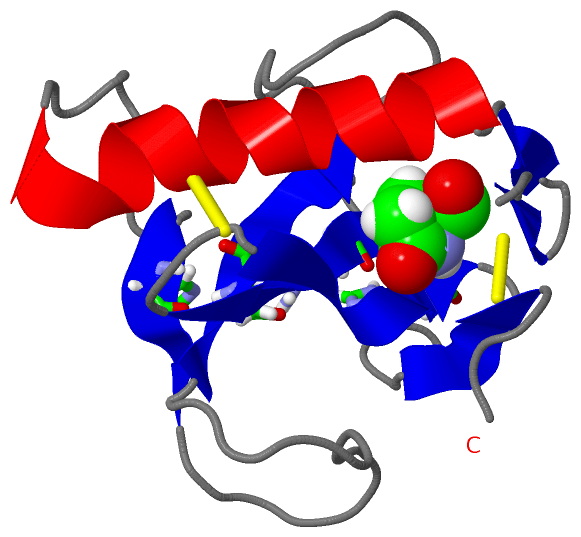
Chains, Units
Summary Information (see also Sequences/Alignments below) |
Ligands, Modified Residues, Ions (1, 1)
NMR Structure (1, 1)
|
Sites (1, 1)
NMR Structure (1, 1)
|
SS Bonds (2, 2)
NMR Structure
|
||||||||||||
Cis Peptide Bonds (3, 3)
NMR Structure
|
||||||||||||||||
SAPs(SNPs)/Variants (0, 0)| (no "SAP(SNP)/Variant" information available for 1RCL) |
PROSITE Motifs (0, 0)| (no "PROSITE Motif" information available for 1RCL) |
Exons (0, 0)| (no "Exon" information available for 1RCL) |
Sequences/Alignments
NMR StructureChain A from PDB Type:PROTEIN Length:106 aligned with RNF1_GIBFU | P10282 from UniProtKB/Swiss-Prot Length:131 Alignment length:106 35 45 55 65 75 85 95 105 115 125 RNF1_GIBFU 26 QSATTCGSTNYSASQVRAAANAACQYYQNDDTAGSSTYPHTYNNYEGFDFPVDGPYQEFPIKSGGVYTGGSPGADRVVINTNCEYAGAITHTGASGNNFVGCSGTN 131 SCOP domains d1rcla_ A: RNase F1 SCOP domains CATH domains -1rclA00 A:2-106 [code=3.10.450.30, no name defined] CATH domains Pfam domains -------------------Ribonuclease-1rclA01 A:20-102 ---- Pfam domains SAPs(SNPs) ---------------------------------------------------------------------------------------------------------- SAPs(SNPs) PROSITE ---------------------------------------------------------------------------------------------------------- PROSITE Transcript ---------------------------------------------------------------------------------------------------------- Transcript 1rcl A 1 xSATTCGSTNYSASQVRAAANAACQYYQNDDTAGSSTYPHTYNNYEGFDFPVDGPYQEFPIKSGGVYTGGSPGADRVVINTNCEYAGAITHTGASGNNFVGCSGTN 106 | 10 20 30 40 50 60 70 80 90 100 | 1-PCA
|
||||||||||||||||||||
SCOP Domains (1, 1)
NMR Structure
 |
CATH Domains (1, 1)
NMR Structure
|
Pfam Domains (1, 1)
NMR Structure
|
Gene Ontology (10, 10)|
NMR Structure(hide GO term definitions) Chain A (RNF1_GIBFU | P10282)
|
||||||||||||||||||||||||||||||||||||||||||||||||||||||||||||||||||||||||
Interactive Views
|
|||||||||||||||||||||||||||||||||||||||||||||||||||||||||||||||||||||||||||||||||||||||||||||||||||||||||||||||||||||||||||||||||||||
Still Images
|
||||||||||||||||
Databases
|
||||||||||||||||||||||||||||||||||||||||||||||||||||||||||||||||||||||||||||||||||||||||||||||||||||||||||||||||||||||||||||||||||||||||||||||||||||||||||||||||
Analysis Tools
|
|||||||||||||||||||||||||||||||||||||||||||||||||||||||||||||
Entries Sharing at Least One Protein Chain (UniProt ID)
Related Entries Specified in the PDB File
|
|