|
|
|
|
 Description
|
|
Compounds
|
||||||||||||||||||||||||||||||||||||||||||||||||||||
Chains, Units
Summary Information (see also Sequences/Alignments below) |
Ligands, Modified Residues, Ions (3, 6)| Asymmetric/Biological Unit (3, 6) |
Sites (6, 6)
Asymmetric Unit (6, 6)
|
SS Bonds (0, 0)| (no "SS Bond" information available for 1R7O) |
Cis Peptide Bonds (2, 2)
Asymmetric/Biological Unit
|
||||||||||||
SAPs(SNPs)/Variants (0, 0)| (no "SAP(SNP)/Variant" information available for 1R7O) |
PROSITE Motifs (1, 1)
Asymmetric/Biological Unit (1, 1)
|
||||||||||||||||||||||||
Exons (0, 0)| (no "Exon" information available for 1R7O) |
Sequences/Alignments
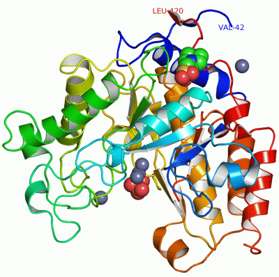
Asymmetric/Biological UnitChain A from PDB Type:PROTEIN Length:353 aligned with MANA_CELJU | P49424 from UniProtKB/Swiss-Prot Length:423 Alignment length:379 51 61 71 81 91 101 111 121 131 141 151 161 171 181 191 201 211 221 231 241 251 261 271 281 291 301 311 321 331 341 351 361 371 381 391 401 411 MANA_CELJU 42 VKPVTVKLVDSQATMETRSLFAFMQEQRRHSIMFGHQHETTQGLTITRTDGTQSDTFNAVGDFAAVYGWDTLSIVAPKAEGDIVAQVKKAYARGGIITVSSHFDNPKTDTQKGVWPVGTSWDQTPAVVDSLPGGAYNPVLNGYLDQVAEWANNLKDEQGRLIPVIFRLYHENTGSWFWWGDKQSTPEQYKQLFRYSVEYLRDVKGVRNFLYAYSPNNFWDVTEANYLERYPGDEWVDVLGFDTYGPVADNADWFRNVVANAALVARMAEARGKIPVISEIGIRAPDIEAGLYDNQWYRKLISGLKADPDAREIAFLLVWRNAPQGVPGPNGTQVPHYWVPANRPENINNGTLEDFQAFYADEFTAFNRDIEQVYQRPTL 420 SCOP domains d1r7oa_ A: Mannanase A, ManA SCOP domains CATH domains 1r7oA00 A:42-420 Glycosidases CATH domains Pfam domains --------Glyco_hydro_26-1r7oA01 A:50-350 ---------------------------------------------------------------------- Pfam domains SAPs(SNPs) ------------------------------------------------------------------------------------------------------------------------------------------------------------------------------------------------------------------------------------------------------------------------------------------------------------------------------------------------------------------------------------------- SAPs(SNPs) PROSITE --------------GH26 PDB: A:56-409 UniProt: 56-409 ----------- PROSITE Transcript ------------------------------------------------------------------------------------------------------------------------------------------------------------------------------------------------------------------------------------------------------------------------------------------------------------------------------------------------------------------------------------------- Transcript 1r7o A 42 VKPVTVKLVDSQATMETRSLFAFMQEQRRHSIMFGHQHETTQGLTITRTDGTQSDTFNAVGDFAAVYGWDTLSIVAPKAEGDIVAQVKKAYARGGIITVSSHFDNPKTDTQKGVWPVGTSWDQTPAVVDSLPGGAYNPVLNGYLDQVAEWANNLKDEQGRLIPVIFRLYHENTGSWFWWGDKQSTPEQYKQLFRYSVEYLRDVKGVRNFLYAYSPNNFWDVTEANYLERYPGDEWVDVLGFDTYGPVADNADWFRNVVANAALVARMAEARGKIPVISEIGIRAPDIEAGLYDNQWYRKLISGLKADPDAREIAFLLVWR--------------------------INNGTLEDFQAFYADEFTAFNRDIEQVYQRPTL 420 51 61 71 81 91 101 111 121 131 141 151 161 171 181 191 201 211 221 231 241 251 261 271 281 291 301 311 321 331 341 351 361 - - |391 401 411 361 388
|
||||||||||||||||||||
SCOP Domains (1, 1)
Asymmetric/Biological Unit
 |
CATH Domains (1, 1)
Asymmetric/Biological Unit
|
Pfam Domains (1, 1)
Asymmetric/Biological Unit
|
Gene Ontology (10, 10)|
Asymmetric/Biological Unit(hide GO term definitions) Chain A (MANA_CELJU | P49424)
|
||||||||||||||||||||||||||||||||||||||||||||||||||||||||||||||||||||||||
Interactive Views
|
|||||||||||||||||||||||||||||||||||||||||||||||||||||||||||||||||||||||||||||||||||||||||||||||||||||||||||||||||||||||||||||||||||||||||||||||||||||||||||||||||||||||||||||||
Still Images
|
||||||||||||||||
Databases
|
||||||||||||||||||||||||||||||||||||||||||||||||||||||||||||||||||||||||||||||||||||||||||||||||||||||||||||||||||||||||||||||||||||||||||||||||||||||||||||||||
Analysis Tools
|
|||||||||||||||||||||||||||||||||||||||||||||||||||||||||||||
Entries Sharing at Least One Protein Chain (UniProt ID)
Related Entries Specified in the PDB File
|
|