Show PDB file:   
         Plain Text   HTML   (compressed file size)
QuickSearch:   
by PDB,NDB,UniProt,PROSITE Code or Search Term(s)  
(-)NMR Structure - model 1
(-)NMR Structure - all models
collapse expand < >
Image NMR Structure - model 1
NMR Structure - model 1  (Jmol Viewer)
Image NMR Structure - all models
NMR Structure - all models  (Jmol Viewer)

(-) Description

Title :  SOLUTION STRUCTURE OF THE DELETION MUTANT DELTA(7-22) OF THE CYTOTOXIC RIBONUCLEASE ALPHA-SARCIN
 
Authors :  M. F. Garcia-Mayoral, L. Garcia-Ortega, M. P. Lillo, J. Santoro, A. Martinez Del Pozo, J. G. Gavilanes, M. Rico, M. Bruix
Date :  09 Oct 03  (Deposition) - 06 Apr 04  (Release) - 24 Feb 09  (Revision)
Method :  SOLUTION NMR
Resolution :  NOT APPLICABLE
Chains :  NMR Structure  :  A  (25x)
Keywords :  Alpha-Beta Protein, Hydrolase (Keyword Search: [Gene Ontology, PubMed, Web (Google))
 
Reference :  M. F. Garcia-Mayoral, L. Garcia-Ortega, M. P. Lillo, J. Santoro, A. Martinez Del Pozo, J. G. Gavilanes, M. Rico, M. Bruix
Nmr Structure Of The Noncytotoxic {Alpha}-Sarcin Mutant {Delta}(7-22): The Importance Of The Native Conformation Of Peripheral Loops For Activity.
Protein Sci. V. 13 1000 2004
PubMed-ID: 15044731  |  Reference-DOI: 10.1110/PS.03532204
(for further references see the PDB file header)

(-) Compounds

Molecule 1 - RIBONUCLEASE ALPHA-SARCIN
    ChainsA
    EC Number3.1.27.10
    EngineeredYES
    Expression SystemESCHERICHIA COLI BL21(DE3)
    Expression System PlasmidPINPGOMPAASD(7-22)
    Expression System StrainBL21(DE3)
    Expression System Taxid469008
    Expression System Vector TypePLASMID
    GeneSAR
    MutationYES
    Organism ScientificASPERGILLUS GIGANTEUS
    Organism Taxid5060
    SynonymRRNA ENDONUCLEASE

 Structural Features

(-) Chains, Units

  
NMR Structure (25x)

Summary Information (see also Sequences/Alignments below)

(-) Ligands, Modified Residues, Ions  (0, 0)

(no "Ligand,Modified Residues,Ions" information available for 1R4Y)

(-) Sites  (0, 0)

(no "Site" information available for 1R4Y)
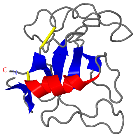
(-) SS Bonds  (2, 2)

NMR Structure
No.Residues
1A:6 -A:134
2A:62 -A:118

(-) Cis Peptide Bonds  (3, 75)

NMR Structure
No.ModelResidues
11, 2, 3, 4, 5, 6, 7, 8, 9, 10, 11, 12, 13, 14, 15, 16, 17, 18, 19, 20, 21, 22, 23, 24, 25Tyr A:34 -Pro A:35
21, 2, 3, 4, 5, 6, 7, 8, 9, 10, 11, 12, 13, 14, 15, 16, 17, 18, 19, 20, 21, 22, 23, 24, 25Lys A:98 -Pro A:99
31, 2, 3, 4, 5, 6, 7, 8, 9, 10, 11, 12, 13, 14, 15, 16, 17, 18, 19, 20, 21, 22, 23, 24, 25Tyr A:112 -Pro A:113

 Sequence-Structure Mapping

(-) SAPs(SNPs)/Variants  (0, 0)

(no "SAP(SNP)/Variant" information available for 1R4Y)

(-) PROSITE Motifs  (0, 0)

(no "PROSITE Motif" information available for 1R4Y)

(-) Exons   (0, 0)

(no "Exon" information available for 1R4Y)

(-) Sequences/Alignments

NMR Structure
   Reformat: Number of residues per line =  ('0' or empty: single-line sequence representation)
  Number of residues per labelling interval =   
  UniProt sequence: complete  aligned part    
   Show mapping: SCOP domains CATH domains Pfam domains Secondary structure (by author)
SAPs(SNPs) PROSITE motifs Exons
(details for a mapped element are shown in a popup box when the mouse pointer rests over it)
Chain A from PDB  Type:PROTEIN  Length:136
 aligned with RNAS_ASPGI | P00655 from UniProtKB/Swiss-Prot  Length:177

    Alignment length:150
                                    37        47        57        67        77        87        97       107       117       127       137       147       157       167       177
           RNAS_ASPGI    28 AVTWTCLNDQKNPKTNKYETKRLLYNQNKAESNSHHAPLSDGKTGSSYPHWFTNGYDGDGKLPKGRTPIKFGKSDCDRPPKHSKDGNGKTDHYLLEFPTFPDGHDYKFDSKKPKENPGPARVIYTYPNKVFCGIIAHTKENQGELKLCSH 177
               SCOP domains d1r4ya_               A: alpha-Sarcin                                                                                                                  SCOP domains
               CATH domains 1r4yA00               A:1-136  [code=3.10.450.30, no name defined]                                                                                     CATH domains
               Pfam domains -------------------------Ribonuclease-1r4yA01 A:12-134                                                                                              -- Pfam domains
         Sec.struct. author ..eee...--------------.eeehhhhhhhhhhh............ee...........................................eeee......................eeee........eeee.........ee... Sec.struct. author
                 SAPs(SNPs) ------------------------------------------------------------------------------------------------------------------------------------------------------ SAPs(SNPs)
                    PROSITE ------------------------------------------------------------------------------------------------------------------------------------------------------ PROSITE
                 Transcript ------------------------------------------------------------------------------------------------------------------------------------------------------ Transcript
                 1r4y A   1 AVTWTCGG--------------LLYNQNKAESNSHHAPLSDGKTGSSYPHWFTNGYDGDGKLPKGRTPIKFGKSDCDRPPKHSKDGNGKTDHYLLEFPTFPDGHDYKFDSKKPKENPGPARVIYTYPNKVFCGIIAHTKENQGELKLCSH 136
                                   | -         -  |     16        26        36        46        56        66        76        86        96       106       116       126       136
                                   8              9                                                                                                                               

   Legend:   → Mismatch (orange background)
  - → Gap (green background, '-', border residues have a numbering label)
    → Modified Residue (blue background, lower-case, 'x' indicates undefined single-letter code, labelled with number + name)
  x → Chemical Group (purple background, 'x', labelled with number + name, e.g. ACE or NH2)
  extra numbering lines below/above indicate numbering irregularities and modified residue names etc., number ends below/above '|'

 Classification and Annotation

(-) SCOP Domains  (1, 1)

NMR Structure

(-) CATH Domains  (1, 1)

NMR Structure
(-)
Class: Alpha Beta (26913)

(-) Pfam Domains  (1, 1)

NMR Structure

(-) Gene Ontology  (11, 11)

NMR Structure(hide GO term definitions)
Chain A   (RNAS_ASPGI | P00655)
molecular function
    GO:0003723    RNA binding    Interacting selectively and non-covalently with an RNA molecule or a portion thereof.
    GO:0004521    endoribonuclease activity    Catalysis of the hydrolysis of ester linkages within ribonucleic acid by creating internal breaks.
    GO:0016787    hydrolase activity    Catalysis of the hydrolysis of various bonds, e.g. C-O, C-N, C-C, phosphoric anhydride bonds, etc. Hydrolase is the systematic name for any enzyme of EC class 3.
    GO:0004518    nuclease activity    Catalysis of the hydrolysis of ester linkages within nucleic acids.
    GO:0033902    rRNA endonuclease activity    Catalysis of the hydrolysis of the phosphodiester linkage between guanosine and adenosine residues at one specific position in 28S rRNA from rat ribosomes.
    GO:0004540    ribonuclease activity    Catalysis of the hydrolysis of phosphodiester bonds in chains of RNA.
biological process
    GO:0090501    RNA phosphodiester bond hydrolysis    The RNA metabolic process in which the phosphodiester bonds between ribonucleotides are cleaved by hydrolysis.
    GO:0090502    RNA phosphodiester bond hydrolysis, endonucleolytic    The chemical reactions and pathways involving the hydrolysis of internal 3',5'-phosphodiester bonds in one or two strands of ribonucleotides.
    GO:0017148    negative regulation of translation    Any process that stops, prevents, or reduces the frequency, rate or extent of the chemical reactions and pathways resulting in the formation of proteins by the translation of mRNA or circRNA.
    GO:0090305    nucleic acid phosphodiester bond hydrolysis    The nucleic acid metabolic process in which the phosphodiester bonds between nucleotides are cleaved by hydrolysis.
cellular component
    GO:0005576    extracellular region    The space external to the outermost structure of a cell. For cells without external protective or external encapsulating structures this refers to space outside of the plasma membrane. This term covers the host cell environment outside an intracellular parasite.

 Visualization

(-) Interactive Views

NMR Structure
  Complete Structure
    Jena3D(integrated viewing of ligand, site, SAP, PROSITE, SCOP information)
    WebMol | AstexViewer[tm]@PDBe
(Java Applets, require no local installation except for Java; loading may be slow)
    STRAP
(Java WebStart application, automatic local installation, requires Java; full application with system access!)
    RasMol
(require local installation)
    Molscript (VRML)
(requires installation of a VRML viewer; select preferred view via VRML and generate a mono or stereo PDF format file)
 
  Ligands, Modified Residues, Ions
(no "Ligands, Modified Residues, Ions" information available for 1r4y)
 
  Sites
(no "Sites" information available for 1r4y)
 
  Cis Peptide Bonds
    Lys A:98 - Pro A:99   [ RasMol ]  
    Tyr A:112 - Pro A:113   [ RasMol ]  
    Tyr A:34 - Pro A:35   [ RasMol ]  
 

(-) Still Images

Jmol
  protein: cartoon or spacefill or dots and stick; nucleic acid: cartoon and stick; ligands: spacefill; active site: stick
Molscript
  protein, nucleic acid: cartoon; ligands: spacefill; active site: ball and stick

 Databases and Analysis Tools

(-) Databases

Access by PDB/NDB ID
  1r4y
    Family and Domain InformationProDom | SYSTERS
    General Structural InformationGlycoscienceDB | MMDB | NDB | OCA | PDB | PDBe | PDBj | PDBsum | PDBWiki | PQS | PROTEOPEDIA
    Orientation in MembranesOPM
    Protein SurfaceSURFACE
    Secondary StructureDSSP (structure derived) | HSSP (homology derived)
    Structural GenomicsGeneCensus
    Structural NeighboursCE | VAST
    Structure ClassificationCATH | Dali | SCOP
    Validation and Original DataBMRB Data View | BMRB Restraints Grid | EDS | PROCHECK | RECOORD | WHAT_CHECK
 
Access by UniProt ID/Accession number
  RNAS_ASPGI | P00655
    Comparative Protein Structure ModelsModBase
    Genomic InformationEnsembl
    Protein-protein InteractionDIP
    Sequence, Family and Domain InformationInterPro | Pfam | SMART | UniProtKB/SwissProt
 
Access by Enzyme Classificator   (EC Number)
  3.1.27.10
    General Enzyme InformationBRENDA | EC-PDB | Enzyme | IntEnz
    PathwayKEGG | MetaCyc
 
Access by Disease Identifier   (MIM ID)
  (no 'MIM ID' available)
    Disease InformationOMIM
 
Access by GenAge ID
  (no 'GenAge ID' available)
    Age Related InformationGenAge

(-) Analysis Tools

Access by PDB/NDB ID
    Domain InformationXDom
    Interatomic Contacts of Structural UnitsCSU
    Ligand-protein ContactsLPC
    Protein CavitiescastP
    Sequence and Secondary StructurePDBCartoon
    Structure AlignmentSTRAP(Java WebStart application, automatic local installation, requires Java; full application with system access!)
    Structure and Sequence BrowserSTING
 
Access by UniProt ID/Accession number
  RNAS_ASPGI | P00655
    Protein Disorder PredictionDisEMBL | FoldIndex | GLOBPLOT (for more information see DisProt)

 Related Entries

(-) Entries Sharing at Least One Protein Chain (UniProt ID)

UniProtKB/Swiss-Prot
        RNAS_ASPGI | P006551de3

(-) Related Entries Specified in the PDB File

1de3 THE WILD TYPE PROTEIN