Show PDB file:   
         Plain Text   HTML   (compressed file size)
QuickSearch:   
by PDB,NDB,UniProt,PROSITE Code or Search Term(s)  
(-)Asym./Biol. Unit
(-)Asym./Biol. Unit - sites
collapse expand < >
Image Asym./Biol. Unit
Asym./Biol. Unit  (Jmol Viewer)
Image Asym./Biol. Unit - sites
Asym./Biol. Unit - sites  (Jmol Viewer)

(-) Description

Title :  1.9A CRYSTAL STRUCTURE OF PROTEIN AQ328 FROM AQUIFEX AEOLICUS
 
Authors :  Y. Qiu, V. Tereshko, Y. Kim, R. Zhang, F. Collart, A. Joachimiak, A. Koss Midwest Center For Structural Genomics (Mcsg)
Date :  08 Oct 03  (Deposition) - 30 Mar 04  (Release) - 13 Jul 11  (Revision)
Method :  X-RAY DIFFRACTION
Resolution :  1.90
Chains :  Asym./Biol. Unit :  A
Keywords :  Structural Genomics, All-Alpha, Histon Fold, Psi, Protein Structure Initiative, Midwest Center For Structural Genomics, Mcsg, Unknown Function (Keyword Search: [Gene Ontology, PubMed, Web (Google))
 
Reference :  Y. Qiu, V. Tereshko, Y. Kim, R. Zhang, F. Collart, M. Yousef, A. Kossiakoff, A. Joachimiak
The Crystal Structure Of Aq_328 From The Hyperthermophilic Bacteria Aquifex Aeolicus Shows An Ancestral Histone Fold.
Proteins V. 62 8 2006
PubMed-ID: 16287087  |  Reference-DOI: 10.1002/PROT.20590

(-) Compounds

Molecule 1 - HYPOTHETICAL PROTEIN AQ_328
    ChainsA
    EngineeredYES
    Expression SystemESCHERICHIA COLI BL21
    Expression System StrainBL21
    Expression System Taxid511693
    GeneAQ_328
    Organism ScientificAQUIFEX AEOLICUS
    Organism Taxid224324
    StrainVF5

 Structural Features

(-) Chains, Units

  1
Asymmetric/Biological Unit A

Summary Information (see also Sequences/Alignments below)

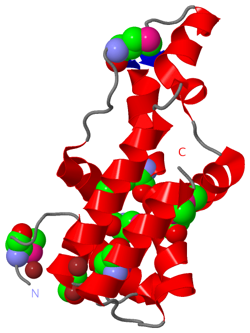
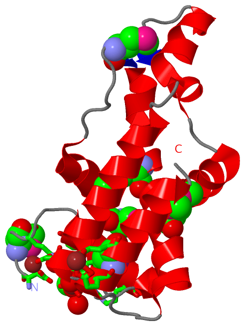
(-) Ligands, Modified Residues, Ions  (3, 10)

Asymmetric/Biological Unit (3, 10)
No.NameCountTypeFull Name
1CAC1Ligand/IonCACODYLATE ION
2MSE6Mod. Amino AcidSELENOMETHIONINE
3ZN3Ligand/IonZINC ION

(-) Sites  (4, 4)

Asymmetric Unit (4, 4)
No.NameEvidenceResiduesDescription
1AC1SOFTWAREHIS A:34 , GLU A:158 , GLU A:161 , CAC A:203BINDING SITE FOR RESIDUE ZN A 201
2AC2SOFTWAREASP A:33 , CAC A:203 , HOH A:514 , HOH A:518 , HOH A:520BINDING SITE FOR RESIDUE ZN A 202
3AC3SOFTWAREARG A:25 , ASP A:30 , ASP A:33 , HIS A:34 , ARG A:37 , GLU A:158 , GLU A:161 , ZN A:201 , ZN A:202 , HOH A:352 , HOH A:514 , HOH A:521BINDING SITE FOR RESIDUE CAC A 203
4AC4SOFTWAREGLU A:21 , ARG A:25 , ASP A:46 , HOH A:452BINDING SITE FOR RESIDUE ZN A 204

(-) SS Bonds  (0, 0)

(no "SS Bond" information available for 1R4V)

(-) Cis Peptide Bonds  (0, 0)

(no "Cis Peptide Bond" information available for 1R4V)

 Sequence-Structure Mapping

(-) SAPs(SNPs)/Variants  (0, 0)

(no "SAP(SNP)/Variant" information available for 1R4V)

(-) PROSITE Motifs  (0, 0)

(no "PROSITE Motif" information available for 1R4V)

(-) Exons   (0, 0)

(no "Exon" information available for 1R4V)

(-) Sequences/Alignments

Asymmetric/Biological Unit
   Reformat: Number of residues per line =  ('0' or empty: single-line sequence representation)
  Number of residues per labelling interval =   
  UniProt sequence: complete  aligned part    
   Show mapping: SCOP domains CATH domains Pfam domains Secondary structure (by author)
SAPs(SNPs) PROSITE motifs Exons
(details for a mapped element are shown in a popup box when the mouse pointer rests over it)
Chain A from PDB  Type:PROTEIN  Length:151
 aligned with Y328_AQUAE | O66665 from UniProtKB/Swiss-Prot  Length:171

    Alignment length:151
                                    30        40        50        60        70        80        90       100       110       120       130       140       150       160       170 
           Y328_AQUAE    21 ETMLRPKGFDKLDHYFRTELDIDLTDETIELLLNSVKAAFGKLFYGAEQRARWNGRDFIALADLNITKALEEHIKNFQKIEQDMGVDELLEYIAFIPPVEMNVGEDLKSEYRNIMGGLLLMHADVIKKATGERKPSREAMEFVAQIVDKVF 171
               SCOP domains d1r4va_ A: Hypothetical protein Aq_328                                                                                                                  SCOP domains
               CATH domains 1r4vA00 A:21-171 Histone, subunit A                                                                                                                     CATH domains
               Pfam domains -----------DUF1931-1r4vA01 A:32-168                                                                                                                 --- Pfam domains
         Sec.struct. author .......hhhhhhhhhhhhhh...hhhhhhhhhhhhhhhhhhh..hhhhhhhhh...eehhhhh..hhhhhhhhhhhhh.....hhhhhhhhhh.......eehhhhhhhhhhhhhhhhhhhhhhhhhhh.....hhhhhhhhhhhhhh.. Sec.struct. author
                 SAPs(SNPs) ------------------------------------------------------------------------------------------------------------------------------------------------------- SAPs(SNPs)
                    PROSITE ------------------------------------------------------------------------------------------------------------------------------------------------------- PROSITE
                 Transcript ------------------------------------------------------------------------------------------------------------------------------------------------------- Transcript
                 1r4v A  21 ETmLRPKGFDKLDHYFRTELDIDLTDETIELLLNSVKAAFGKLFYGAEQRARWNGRDFIALADLNITKALEEHIKNFQKIEQDmGVDELLEYIAFIPPVEmNVGEDLKSEYRNImGGLLLmHADVIKKATGERKPSREAmEFVAQIVDKVF 171
                              |     30        40        50        60        70        80        90       100   |   110       120|      130    |  140|      150       160       170 
                              |                                                                              104-MSE          121-MSE       135-MSE |                160-MSE       
                             23-MSE                                                                                                               141-MSE                          

   Legend:   → Mismatch (orange background)
  - → Gap (green background, '-', border residues have a numbering label)
    → Modified Residue (blue background, lower-case, 'x' indicates undefined single-letter code, labelled with number + name)
  x → Chemical Group (purple background, 'x', labelled with number + name, e.g. ACE or NH2)
  extra numbering lines below/above indicate numbering irregularities and modified residue names etc., number ends below/above '|'

 Classification and Annotation

(-) SCOP Domains  (1, 1)

Asymmetric/Biological Unit

(-) CATH Domains  (1, 1)

Asymmetric/Biological Unit

(-) Pfam Domains  (1, 1)

Asymmetric/Biological Unit

(-) Gene Ontology  (1, 1)

Asymmetric/Biological Unit(hide GO term definitions)
Chain A   (Y328_AQUAE | O66665)
molecular function
    GO:0046982    protein heterodimerization activity    Interacting selectively and non-covalently with a nonidentical protein to form a heterodimer.

 Visualization

(-) Interactive Views

Asymmetric/Biological Unit
  Complete Structure
    Jena3D(integrated viewing of ligand, site, SAP, PROSITE, SCOP information)
    WebMol | AstexViewer[tm]@PDBe
(Java Applets, require no local installation except for Java; loading may be slow)
    STRAP
(Java WebStart application, automatic local installation, requires Java; full application with system access!)
    RasMol
(require local installation)
    Molscript (VRML)
(requires installation of a VRML viewer; select preferred view via VRML and generate a mono or stereo PDF format file)
 
  Ligands, Modified Residues, Ions
    CAC  [ RasMol | Jena3D ]  +environment [ RasMol | Jena3D ]
    MSE  [ RasMol | Jena3D ]  +environment [ RasMol | Jena3D ]
    ZN  [ RasMol | Jena3D ]  +environment [ RasMol | Jena3D ]
 
  Sites
    AC1  [ RasMol ]  +environment [ RasMol ]
    AC2  [ RasMol ]  +environment [ RasMol ]
    AC3  [ RasMol ]  +environment [ RasMol ]
    AC4  [ RasMol ]  +environment [ RasMol ]
 
  Cis Peptide Bonds
(no "Cis Peptide Bonds" information available for 1r4v)
 

(-) Still Images

Jmol
  protein: cartoon or spacefill or dots and stick; nucleic acid: cartoon and stick; ligands: spacefill; active site: stick
Molscript
  protein, nucleic acid: cartoon; ligands: spacefill; active site: ball and stick

 Databases and Analysis Tools

(-) Databases

Access by PDB/NDB ID
  1r4v
    Family and Domain InformationProDom | SYSTERS
    General Structural InformationGlycoscienceDB | MMDB | NDB | OCA | PDB | PDBe | PDBj | PDBsum | PDBWiki | PQS | PROTEOPEDIA
    Orientation in MembranesOPM
    Protein SurfaceSURFACE
    Secondary StructureDSSP (structure derived) | HSSP (homology derived)
    Structural GenomicsGeneCensus
    Structural NeighboursCE | VAST
    Structure ClassificationCATH | Dali | SCOP
    Validation and Original DataBMRB Data View | BMRB Restraints Grid | EDS | PROCHECK | RECOORD | WHAT_CHECK
 
Access by UniProt ID/Accession number
  Y328_AQUAE | O66665
    Comparative Protein Structure ModelsModBase
    Genomic InformationEnsembl
    Protein-protein InteractionDIP
    Sequence, Family and Domain InformationInterPro | Pfam | SMART | UniProtKB/SwissProt
 
Access by Enzyme Classificator   (EC Number)
  (no 'Enzyme Classificator' available)
    General Enzyme InformationBRENDA | EC-PDB | Enzyme | IntEnz
    PathwayKEGG | MetaCyc
 
Access by Disease Identifier   (MIM ID)
  (no 'MIM ID' available)
    Disease InformationOMIM
 
Access by GenAge ID
  (no 'GenAge ID' available)
    Age Related InformationGenAge

(-) Analysis Tools

Access by PDB/NDB ID
    Domain InformationXDom
    Interatomic Contacts of Structural UnitsCSU
    Ligand-protein ContactsLPC
    Protein CavitiescastP
    Sequence and Secondary StructurePDBCartoon
    Structure AlignmentSTRAP(Java WebStart application, automatic local installation, requires Java; full application with system access!)
    Structure and Sequence BrowserSTING
 
Access by UniProt ID/Accession number
  Y328_AQUAE | O66665
    Protein Disorder PredictionDisEMBL | FoldIndex | GLOBPLOT (for more information see DisProt)

 Related Entries

(-) Entries Sharing at Least One Protein Chain (UniProt ID)

(no "Entries Sharing at Least One Protein Chain" available for 1R4V)

(-) Related Entries Specified in the PDB File

(no "Related Entries Specified in the PDB File" available for 1R4V)