Show PDB file:   
         Plain Text   HTML   (compressed file size)
QuickSearch:   
by PDB,NDB,UniProt,PROSITE Code or Search Term(s)  
(-)Biol.Unit 1 - manually
(-)Asym.Unit - manually
(-)Asymmetric Unit
(-)Asym. Unit - sites
(-)Biological Unit 1
(-)Biol. Unit 1 - sites
collapse expand < >
Image Biol.Unit 1 - manually
Biol.Unit 1 - manually  (Jmol Viewer)
Image Asym.Unit - manually
Asym.Unit - manually  (Jmol Viewer)
Image Asymmetric Unit
Asymmetric Unit  (Jmol Viewer)
Image Asym. Unit - sites
Asym. Unit - sites  (Jmol Viewer)
Image Biological Unit 1
Biological Unit 1  (Jmol Viewer)
Image Biol. Unit 1 - sites
Biol. Unit 1 - sites  (Jmol Viewer)

(-) Description

Title :  CRYSTAL STRUCTURE OF A HUMAN MITOCHONDRIAL FERRITIN
 
Authors :  B. Corsi, P. Santambrogio, P. Arosio, S. Levi, B. Langlois D'Estainto T. Granier, B. Gallois, J. M. Chevallier, G. Precigoux
Date :  19 Sep 03  (Deposition) - 29 Jun 04  (Release) - 13 Jul 11  (Revision)
Method :  X-RAY DIFFRACTION
Resolution :  1.70
Chains :  Asym. Unit :  A
Biol. Unit 1:  A  (24x)
Keywords :  Iron Storage; Ferritin; X-Ray Crystallography, Metal Binding Protein (Keyword Search: [Gene Ontology, PubMed, Web (Google))
 
Reference :  B. Langlois D'Estaintot, P. Santambrogio, T. Granier, B. Gallois, J. M. Chevallier, G. Precigoux, S. Levi, P. Arosio
Crystal Structure And Biochemical Properties Of The Human Mitochondrial Ferritin And Its Mutant Ser144Ala
J. Mol. Biol. V. 340 277 2004
PubMed-ID: 15201052  |  Reference-DOI: 10.1016/J.JMB.2004.04.036
(for further references see the PDB file header)

(-) Compounds

Molecule 1 - MITOCHONDRIAL FERRITIN
    ChainsA
    EngineeredYES
    Expression SystemESCHERICHIA COLI
    Expression System Taxid562
    Expression System Vector TypePLASMID
    Organism CommonHUMAN
    Organism ScientificHOMO SAPIENS
    Organism Taxid9606
    SynonymFERRITIN HEAVY CHAIN;
FERRITIN H SUBUNIT

 Structural Features

(-) Chains, Units

  1
Asymmetric Unit A
Biological Unit 1 (24x)A

Summary Information (see also Sequences/Alignments below)

(-) Ligands, Modified Residues, Ions  (1, 5)

Asymmetric Unit (1, 5)
No.NameCountTypeFull Name
1MG5Ligand/IonMAGNESIUM ION
Biological Unit 1 (0, 0)
No.NameCountTypeFull Name
1MG-1Ligand/IonMAGNESIUM ION

(-) Sites  (5, 5)

Asymmetric Unit (5, 5)
No.NameEvidenceResiduesDescription
1AC1SOFTWAREGLU A:27 , GLU A:62 , HIS A:65 , HOH A:636 , HOH A:637BINDING SITE FOR RESIDUE MG A 301
2AC2SOFTWAREHIS A:173 , HOH A:516 , HOH A:638BINDING SITE FOR RESIDUE MG A 302
3AC3SOFTWAREHOH A:639 , HOH A:640 , HOH A:641 , HOH A:642 , HOH A:643 , HOH A:644BINDING SITE FOR RESIDUE MG A 305
4AC4SOFTWAREHOH A:645 , HOH A:646 , HOH A:647 , HOH A:648 , HOH A:649 , HOH A:650BINDING SITE FOR RESIDUE MG A 306
5AC5SOFTWAREASP A:131 , GLU A:134 , HOH A:651 , HOH A:652 , HOH A:653 , HOH A:654BINDING SITE FOR RESIDUE MG A 310

(-) SS Bonds  (0, 0)

(no "SS Bond" information available for 1R03)

(-) Cis Peptide Bonds  (1, 1)

Asymmetric Unit
No.Residues
1Ala A:160 -Pro A:161

 Sequence-Structure Mapping

(-) SAPs(SNPs)/Variants  (0, 0)

(no "SAP(SNP)/Variant" information available for 1R03)

(-) PROSITE Motifs  (2, 2)

Asymmetric Unit (2, 2)
 PROSITEUniProtKBPDB
No.IDACDescriptionIDLocationCountLocation
1FERRITIN_LIKEPS50905 Ferritin-like diiron domain profile.FTMT_HUMAN70-219  1A:10-159
2FERRITIN_2PS00204 Ferritin iron-binding regions signature 2.FTMT_HUMAN186-206  1A:126-146
Biological Unit 1 (2, 48)
 PROSITEUniProtKBPDB
No.IDACDescriptionIDLocationCountLocation
1FERRITIN_LIKEPS50905 Ferritin-like diiron domain profile.FTMT_HUMAN70-219  24A:10-159
2FERRITIN_2PS00204 Ferritin iron-binding regions signature 2.FTMT_HUMAN186-206  24A:126-146

(-) Exons   (1, 1)

Asymmetric Unit (1, 1)
 ENSEMBLUniProtKBPDB
No.Transcript IDExonExon IDGenome LocationLengthIDLocationLengthCountLocationLength
1.1ENST000003213391ENSE00001262353chr5:121187650-121188519870FTMT_HUMAN1-2452451A:6-176171

(-) Sequences/Alignments

Asymmetric Unit
   Reformat: Number of residues per line =  ('0' or empty: single-line sequence representation)
  Number of residues per labelling interval =   
  UniProt sequence: complete  aligned part    
   Show mapping: SCOP domains CATH domains Pfam domains Secondary structure (by author)
SAPs(SNPs) PROSITE motifs Exons
(details for a mapped element are shown in a popup box when the mouse pointer rests over it)
Chain A from PDB  Type:PROTEIN  Length:171
 aligned with FTMT_HUMAN | Q8N4E7 from UniProtKB/Swiss-Prot  Length:242

    Alignment length:171
                                    75        85        95       105       115       125       135       145       155       165       175       185       195       205       215       225       235 
           FTMT_HUMAN    66 SRVRQNFHPDSEAAINRQINLELYASYVYLSMAYYFSRDDVALNNFSRYFLHQSREETEHAEKLMRLQNQRGGRIRLQDIKKPEQDDWESGLHAMECALLLEKNVNQSLLELHALASDKGDPHLCDFLETYYLNEQVKSIKELGDHVHNLVKMGAPDAGLAEYLFDTHTLG 236
               SCOP domains d1r03a_ A: (Apo)ferritin                                                                                                                                                    SCOP domains
               CATH domains 1r03A00 A:6-176  [code=1.20.1260.10, no name defined]                                                                                                                       CATH domains
               Pfam domains -----------Ferritin-1r03A01 A:17-158                                                                                                                     ------------------ Pfam domains
         Sec.struct. author .......hhhhhhhhhhhhhhhhhhhhhhhhhhhhh......hhhhhhhhhhhhhhhhhhhhhhhhhhhhhh.................hhhhhhhhhhhhhhhhh.hhhhhhhhhhhh.hhhhhhhhhhhhhhhhhhhhhhhhhhhhhhhhhh.....hhhhhhhhhh.. Sec.struct. author
                 SAPs(SNPs) --------------------------------------------------------------------------------------------------------------------------------------------------------------------------- SAPs(SNPs)
                PROSITE (1) ----FERRITIN_LIKE  PDB: A:10-159 UniProt: 70-219                                                                                                          ----------------- PROSITE (1)
                PROSITE (2) ------------------------------------------------------------------------------------------------------------------------FERRITIN_2           ------------------------------ PROSITE (2)
               Transcript 1 Exon 1.1  PDB: A:6-176 UniProt: 1-245 [INCOMPLETE]                                                                                                                          Transcript 1
                 1r03 A   6 SRVRQNFHPDSEAAINRQINLELYASYVYLSMAYYFSRDDVALNNFSRYFLHQSREETEHAEKLMRLQNQRGGRIRLQDIKKPEQDDWESGLHAMECALLLEKNVNQSLLELHALASDKGDPHLCDFLETYYLNEQVKSIKELGDHVHNLVKMGAPDAGLAEYLFDTHTLG 176
                                    15        25        35        45        55        65        75        85        95       105       115       125       135       145       155       165       175 

   Legend:   → Mismatch (orange background)
  - → Gap (green background, '-', border residues have a numbering label)
    → Modified Residue (blue background, lower-case, 'x' indicates undefined single-letter code, labelled with number + name)
  x → Chemical Group (purple background, 'x', labelled with number + name, e.g. ACE or NH2)
  extra numbering lines below/above indicate numbering irregularities and modified residue names etc., number ends below/above '|'

 Classification and Annotation

(-) SCOP Domains  (1, 1)

Asymmetric Unit

(-) CATH Domains  (1, 1)

Asymmetric Unit

(-) Pfam Domains  (1, 1)

Asymmetric Unit
(-)
Clan: Ferritin (185)

(-) Gene Ontology  (13, 13)

Asymmetric Unit(hide GO term definitions)
Chain A   (FTMT_HUMAN | Q8N4E7)
molecular function
    GO:0008199    ferric iron binding    Interacting selectively and non-covalently with ferric iron, Fe(III).
    GO:0004322    ferroxidase activity    Catalysis of the reaction: 4 Fe2+ + 4 H+ + O2 = 4 Fe3+ + 2 H2O.
    GO:0046872    metal ion binding    Interacting selectively and non-covalently with any metal ion.
    GO:0016491    oxidoreductase activity    Catalysis of an oxidation-reduction (redox) reaction, a reversible chemical reaction in which the oxidation state of an atom or atoms within a molecule is altered. One substrate acts as a hydrogen or electron donor and becomes oxidized, while the other acts as hydrogen or electron acceptor and becomes reduced.
biological process
    GO:0006879    cellular iron ion homeostasis    Any process involved in the maintenance of an internal steady state of iron ions at the level of a cell.
    GO:0006826    iron ion transport    The directed movement of iron (Fe) ions into, out of or within a cell, or between cells, by means of some agent such as a transporter or pore.
    GO:0055114    oxidation-reduction process    A metabolic process that results in the removal or addition of one or more electrons to or from a substance, with or without the concomitant removal or addition of a proton or protons.
    GO:1904234    positive regulation of aconitate hydratase activity    Any process that activates or increases the frequency, rate or extent of aconitate hydratase activity.
    GO:0008284    positive regulation of cell proliferation    Any process that activates or increases the rate or extent of cell proliferation.
    GO:0051349    positive regulation of lyase activity    Any process that activates or increases the frequency, rate or extent of lyase activity, the catalysis of the cleavage of C-C, C-O, C-N and other bonds by other means than by hydrolysis or oxidation, or conversely adding a group to a double bond.
    GO:1904231    positive regulation of succinate dehydrogenase activity    Any process that activates or increases the frequency, rate or extent of succinate dehydrogenase activity.
cellular component
    GO:0005739    mitochondrion    A semiautonomous, self replicating organelle that occurs in varying numbers, shapes, and sizes in the cytoplasm of virtually all eukaryotic cells. It is notably the site of tissue respiration.
    GO:0005634    nucleus    A membrane-bounded organelle of eukaryotic cells in which chromosomes are housed and replicated. In most cells, the nucleus contains all of the cell's chromosomes except the organellar chromosomes, and is the site of RNA synthesis and processing. In some species, or in specialized cell types, RNA metabolism or DNA replication may be absent.
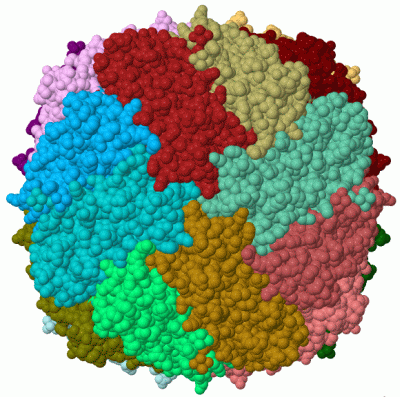
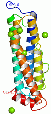
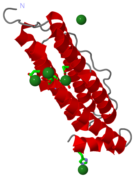
 Visualization

(-) Interactive Views

Asymmetric Unit
  Complete Structure
    Jena3D(integrated viewing of ligand, site, SAP, PROSITE, SCOP information)
    WebMol | AstexViewer[tm]@PDBe
(Java Applets, require no local installation except for Java; loading may be slow)
    STRAP
(Java WebStart application, automatic local installation, requires Java; full application with system access!)
    RasMol
(require local installation)
    Molscript (VRML)
(requires installation of a VRML viewer; select preferred view via VRML and generate a mono or stereo PDF format file)
 
  Ligands, Modified Residues, Ions
    MG  [ RasMol | Jena3D ]  +environment [ RasMol | Jena3D ]
 
  Sites
    AC1  [ RasMol ]  +environment [ RasMol ]
    AC2  [ RasMol ]  +environment [ RasMol ]
    AC3  [ RasMol ]  +environment [ RasMol ]
    AC4  [ RasMol ]  +environment [ RasMol ]
    AC5  [ RasMol ]  +environment [ RasMol ]
 
  Cis Peptide Bonds
    Ala A:160 - Pro A:161   [ RasMol ]  
 
Biological Unit
  Complete Structure
    Biological Unit 1  [ Jena3D ]

(-) Still Images

Jmol
  protein: cartoon or spacefill or dots and stick; nucleic acid: cartoon and stick; ligands: spacefill; active site: stick
Molscript
  protein, nucleic acid: cartoon; ligands: spacefill; active site: ball and stick

 Databases and Analysis Tools

(-) Databases

Access by PDB/NDB ID
  1r03
    Family and Domain InformationProDom | SYSTERS
    General Structural InformationGlycoscienceDB | MMDB | NDB | OCA | PDB | PDBe | PDBj | PDBsum | PDBWiki | PQS | PROTEOPEDIA
    Orientation in MembranesOPM
    Protein SurfaceSURFACE
    Secondary StructureDSSP (structure derived) | HSSP (homology derived)
    Structural GenomicsGeneCensus
    Structural NeighboursCE | VAST
    Structure ClassificationCATH | Dali | SCOP
    Validation and Original DataBMRB Data View | BMRB Restraints Grid | EDS | PROCHECK | RECOORD | WHAT_CHECK
 
Access by UniProt ID/Accession number
  FTMT_HUMAN | Q8N4E7
    Comparative Protein Structure ModelsModBase
    Genomic InformationEnsembl
    Protein-protein InteractionDIP
    Sequence, Family and Domain InformationInterPro | Pfam | SMART | UniProtKB/SwissProt
 
Access by Enzyme Classificator   (EC Number)
  (no 'Enzyme Classificator' available)
    General Enzyme InformationBRENDA | EC-PDB | Enzyme | IntEnz
    PathwayKEGG | MetaCyc
 
Access by Disease Identifier   (MIM ID)
  (no 'MIM ID' available)
    Disease InformationOMIM
 
Access by GenAge ID
  (no 'GenAge ID' available)
    Age Related InformationGenAge

(-) Analysis Tools

Access by PDB/NDB ID
    Domain InformationXDom
    Interatomic Contacts of Structural UnitsCSU
    Ligand-protein ContactsLPC
    Protein CavitiescastP
    Sequence and Secondary StructurePDBCartoon
    Structure AlignmentSTRAP(Java WebStart application, automatic local installation, requires Java; full application with system access!)
    Structure and Sequence BrowserSTING
 
Access by UniProt ID/Accession number
  FTMT_HUMAN | Q8N4E7
    Protein Disorder PredictionDisEMBL | FoldIndex | GLOBPLOT (for more information see DisProt)

 Related Entries

(-) Entries Sharing at Least One Protein Chain (UniProt ID)

(no "Entries Sharing at Least One Protein Chain" available for 1R03)

(-) Related Entries Specified in the PDB File

2fha 82% SEQUENCE IDENTITY WITH 2FHA