|
|
|
|
 Description
|
|
Compounds
|
||||||||||||||||||||||||||||||||||||
Chains, Units
Summary Information (see also Sequences/Alignments below) |
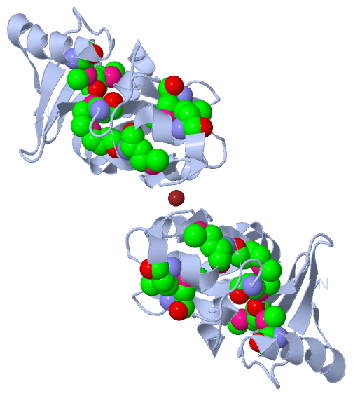
Ligands, Modified Residues, Ions (2, 8)| Asymmetric Unit (2, 8) Biological Unit 1 (1, 7) Biological Unit 2 (1, 14) |
Sites (1, 1)
Asymmetric Unit (1, 1)
|
SS Bonds (0, 0)| (no "SS Bond" information available for 1Q7H) |
Cis Peptide Bonds (0, 0)| (no "Cis Peptide Bond" information available for 1Q7H) |
SAPs(SNPs)/Variants (0, 0)| (no "SAP(SNP)/Variant" information available for 1Q7H) |
PROSITE Motifs (0, 0)| (no "PROSITE Motif" information available for 1Q7H) |
Exons (0, 0)| (no "Exon" information available for 1Q7H) |
Sequences/Alignments
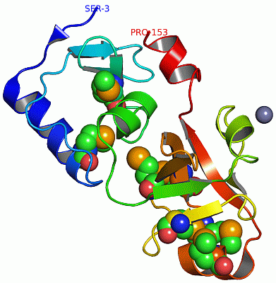
Asymmetric UnitChain A from PDB Type:PROTEIN Length:151 aligned with Q9HIB8_THEAC | Q9HIB8 from UniProtKB/TrEMBL Length:153 Alignment length:151 12 22 32 42 52 62 72 82 92 102 112 122 132 142 152 Q9HIB8_THEAC 3 SKHFISKKEAKRIWEAMARYGIDITGESLEVAAQKSASAYYIGGKPMVFQAGDLIPSVYLLNYRNPSRNIVTVDEGAEPHILNGSDLFAPGIVSMDDSIRKGDMIFVKSSKGYFIAVGMAEMDAGEVMATKRGKAARIIHFPGDELIRAFP 153 SCOP domains d1q7ha2 A:3-68 Hypothetical protein Ta1423, N-terminal domain d1q7ha1 A:69-153 Hypothetical protein Ta1423, C-terminal domain SCOP domains CATH domains 1q7hA01 A:3-67 Pre-PUA domain; domain 1 1q7hA02 A:68-153 [code=2.30.130.10, no name defined] CATH domains Pfam domains DUF1947-1q7hA02 A:3-67 ---PUA-1q7hA01 A:71-144 --------- Pfam domains SAPs(SNPs) ------------------------------------------------------------------------------------------------------------------------------------------------------- SAPs(SNPs) PROSITE ------------------------------------------------------------------------------------------------------------------------------------------------------- PROSITE Transcript ------------------------------------------------------------------------------------------------------------------------------------------------------- Transcript 1q7h A 3 SKHFISKKEAKRIWEQmSRYGIDITGESLEVAAQKSASAYYIGGKPmVFQAGDLIPSVYLLNYRNPSRNIVTVDEGAEPHILNGSDLFAPGIVSmDDSIRKGDmIFVKSSKGYFIAVGmAEmDAGEVmATKRGKAARIIHFPGDELIRAFP 153 12 | 22 32 42 | 52 62 72 82 92 | 102 | 112 122 | 132 142 152 19-MSE 49-MSE 97-MSE 106-MSE 121-MSE | 124-MSE | 130-MSE
|
||||||||||||||||||||
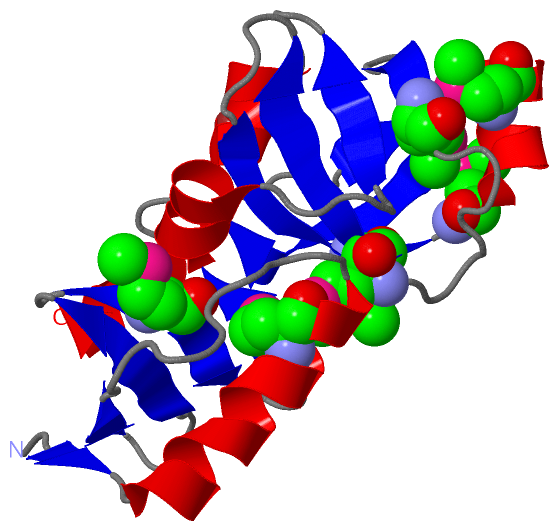
SCOP Domains (2, 2)
Asymmetric Unit
 |
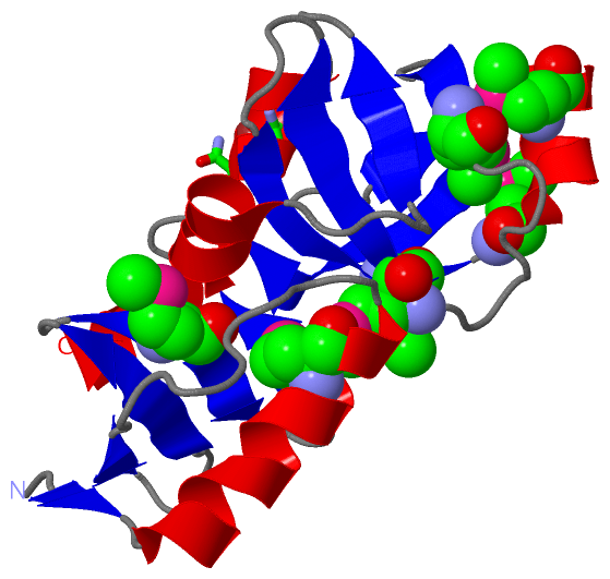
CATH Domains (2, 2)
Asymmetric Unit
|
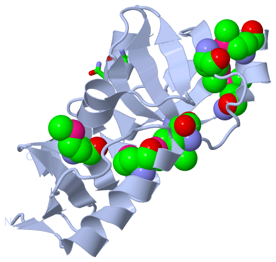
Pfam Domains (2, 2)
Asymmetric Unit
|
Gene Ontology (1, 1)|
Asymmetric Unit(hide GO term definitions) Chain A (Q9HIB8_THEAC | Q9HIB8)
|
||||||||||||
Interactive Views
|
||||||||||||||||||||||||||||||||||||||||||||||||||||||||||||||||||||||||||||||||||||||||||||||||||||||||||||||||||||||||||||||||||||||||||||||||||||
Still Images
|
||||||||||||||||
Databases
|
||||||||||||||||||||||||||||||||||||||||||||||||||||||||||||||||||||||||||||||||||||||||||||||||||||||||||||||||||||||||||||||||||||||||||||||||||||||||||||||||
Analysis Tools
|
|||||||||||||||||||||||||||||||||||||||||||||||||||||||||||||
Entries Sharing at Least One Protein Chain (UniProt ID)
Related Entries Specified in the PDB File
|
|