|
|
|
|
 Description
|
|
Compounds
|
||||||||||||||||||||||||||||||||||||||||||||||||||||||||||||||||||||||||||
Chains, Units
Summary Information (see also Sequences/Alignments below) |
Ligands, Modified Residues, Ions (1, 1)
Asymmetric/Biological Unit (1, 1)
|
Sites (1, 1)
Asymmetric Unit (1, 1)
|
SS Bonds (0, 0)| (no "SS Bond" information available for 1Q2C) |
Cis Peptide Bonds (0, 0)| (no "Cis Peptide Bond" information available for 1Q2C) |
SAPs(SNPs)/Variants (0, 0)| (no "SAP(SNP)/Variant" information available for 1Q2C) |
PROSITE Motifs (0, 0)| (no "PROSITE Motif" information available for 1Q2C) |
Exons (0, 0)| (no "Exon" information available for 1Q2C) |
Sequences/Alignments
Asymmetric/Biological UnitChain A from PDB Type:PROTEIN Length:162 aligned with Q27198_TETTH | Q27198 from UniProtKB/TrEMBL Length:418 Alignment length:162 58 68 78 88 98 108 118 128 138 148 158 168 178 188 198 208 Q27198_TETTH 49 LDFDILTNDGTHRNMKLLIDLKNIFSRQLPKMPKEYIVKLVLDRHHESMVILKNKQKVIGGICFRQYKPQRFAEVAFLAVTANEQVRGYGTRLMNKFKDHMQKQNIEYLLTYADNFAIGYFKKQGFTKEHRMPQEKWKGYIKDYDGGTLMECYIHPYVDYGN 210 SCOP domains d1q2ca_ A: Catalytic domain of GCN5 histone acetyltransferase SCOP domains CATH domains 1q2cA00 A:49-210 [code=3.40.630.30, no name defined] CATH domains Pfam domains ---------------------------------------------------Acetyltransf_1-1q2cA01 A:100-175 ----------------------------------- Pfam domains SAPs(SNPs) ------------------------------------------------------------------------------------------------------------------------------------------------------------------ SAPs(SNPs) PROSITE ------------------------------------------------------------------------------------------------------------------------------------------------------------------ PROSITE Transcript ------------------------------------------------------------------------------------------------------------------------------------------------------------------ Transcript 1q2c A 49 LDFDILTNDGTHRNMKLLIDLKNIFSRQLPKMPKEYIVKLVFDRHHESMVILKNKQKVIGGICFRQYKPQRFAEVAFLAVTANEQVRGYGTRLMNKFKDHMQKQNIEYLLTYADNFAIGYFKKQGFTKEHRMPQEKWKGYIKDYDGGTLMECYIHPYVDYGR 210 58 68 78 88 98 108 118 128 138 148 158 168 178 188 198 208
Chain B from PDB Type:PROTEIN Length:8
SCOP domains -------- SCOP domains
CATH domains -------- CATH domains
Pfam domains -------- Pfam domains
SAPs(SNPs) -------- SAPs(SNPs)
PROSITE -------- PROSITE
Transcript -------- Transcript
1q2c B 308 KGLGKGGA 315
|
||||||||||||||||||||
SCOP Domains (1, 1)
Asymmetric/Biological Unit
 |
CATH Domains (1, 1)
Asymmetric/Biological Unit
|
Pfam Domains (1, 1)
Asymmetric/Biological Unit
|
Gene Ontology (1, 1)|
Asymmetric/Biological Unit(hide GO term definitions) Chain A (Q27198_TETTH | Q27198)
|
||||||||||||
Interactive Views
|
||||||||||||||||||||||||||||||||||||||||||||||||||||||||||||||||||||||||||||||||||||||||||||||||||||||||||||||||||||||
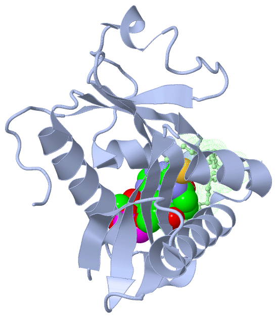
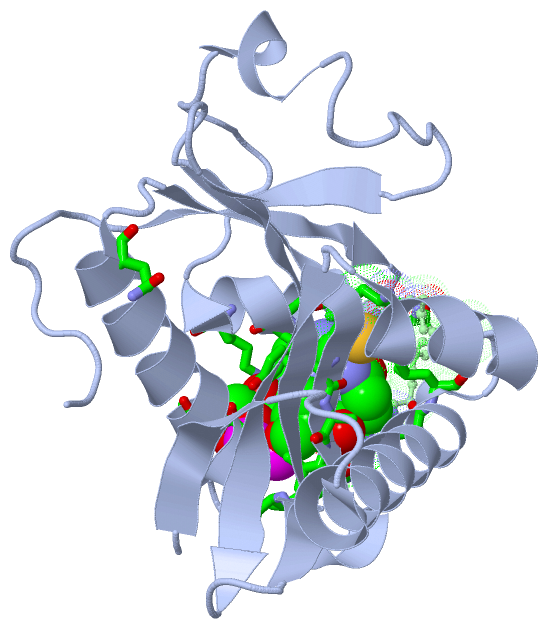
Still Images
|
||||||||||||||||
Databases
|
||||||||||||||||||||||||||||||||||||||||||||||||||||||||||||||||||||||||||||||||||||||||||||||||||||||||||||||||||||||||||||||||||||||||||||||||||||||||||||||||
Analysis Tools
|
|||||||||||||||||||||||||||||||||||||||||||||||||||||||||||||
Entries Sharing at Least One Protein Chain (UniProt ID)
Related Entries Specified in the PDB File
|
|