|
|
|
|
 Description
|
|
Compounds
|
||||||||||||||||||||||||||||||||||||||||||||||||
Chains, Units
Summary Information (see also Sequences/Alignments below) |
Ligands, Modified Residues, Ions (0, 0)| (no "Ligand,Modified Residues,Ions" information available for 1Q27) |
Sites (0, 0)| (no "Site" information available for 1Q27) |
SS Bonds (0, 0)| (no "SS Bond" information available for 1Q27) |
Cis Peptide Bonds (0, 0)| (no "Cis Peptide Bond" information available for 1Q27) |
SAPs(SNPs)/Variants (0, 0)| (no "SAP(SNP)/Variant" information available for 1Q27) |
PROSITE Motifs (2, 2)
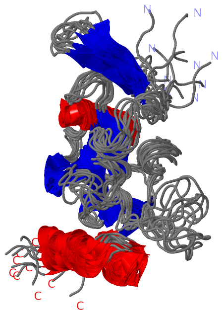
NMR Structure (2, 2)
|
||||||||||||||||||||||||||||||||
Exons (0, 0)| (no "Exon" information available for 1Q27) |
Sequences/Alignments
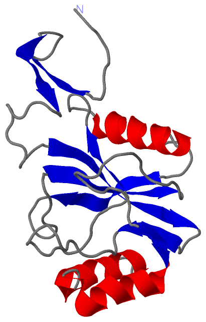
NMR StructureChain A from PDB Type:PROTEIN Length:171 aligned with Y079_DEIRA | Q9RY71 from UniProtKB/Swiss-Prot Length:171 Alignment length:171 10 20 30 40 50 60 70 80 90 100 110 120 130 140 150 160 170 Y079_DEIRA 1 MGGVSDERLDLVNERDEVVGQILRTDPALRWERVRVVNAFLRNSQGQLWIPRRSPSKSLFPNALDVSVGGAVQSGETYEEAFRREAREELNVEIDALSWRPLASFSPFQTTLSSFMCVYELRSDATPIFNPNDISGGEWLTPEHLLARIAAGEAAKGDLAELVRRCYREEE 171 SCOP domains d1q27a_ A: Hypothetical protein DR0079 SCOP domains CATH domains 1q27A00 A:1-171 Nucleoside Triphosphate Pyrophosphohydrolase CATH domains Pfam domains --------------------------------NUDIX-1q27A01 A:33-167 ---- Pfam domains SAPs(SNPs) --------------------------------------------------------------------------------------------------------------------------------------------------------------------------- SAPs(SNPs) PROSITE (1) -------------------------------NUDIX PDB: A:32-162 UniProt: 32-162 --------- PROSITE (1) PROSITE (2) ---------------------------------------------------------------------NUDIX_BOX PDB: A:70-9-------------------------------------------------------------------------------- PROSITE (2) Transcript --------------------------------------------------------------------------------------------------------------------------------------------------------------------------- Transcript 1q27 A 1 MGGVSDERLDLVNERDEVVGQILRTDPALRWERVRVVNAFLRNSQGQLWIPRRSPSKSLFPNALDVSVGGAVQSGETYEEAFRREAREELNVEIDALSWRPLASFSPFQTTLSSFMCVYELRSDATPIFNPNDISGGEWLTPEHLLARIAAGEAAKGDLAELVRRCYREEE 171 10 20 30 40 50 60 70 80 90 100 110 120 130 140 150 160 170
|
||||||||||||||||||||
SCOP Domains (1, 1)
NMR Structure
 |
CATH Domains (1, 1)
NMR Structure
|
Pfam Domains (1, 1)
NMR Structure
|
Gene Ontology (3, 3)|
NMR Structure(hide GO term definitions) Chain A (Y079_DEIRA | Q9RY71)
|
||||||||||||||||||||||||
Interactive Views
|
||||||||||||||||||||||||||||||||||||||||||||||||||||||||||||||||||||||||||||||||||||||||||||||||||||||||||||||||||||
Still Images
|
||||||||||||||||
Databases
|
||||||||||||||||||||||||||||||||||||||||||||||||||||||||||||||||||||||||||||||||||||||||||||||||||||||||||||||||||||||||||||||||||||||||||||||||||||||||||||||||
Analysis Tools
|
|||||||||||||||||||||||||||||||||||||||||||||||||||||||||||||
Entries Sharing at Least One Protein Chain (UniProt ID)
Related Entries Specified in the PDB File
|
|