Show PDB file:   
         Plain Text   HTML   (compressed file size)
QuickSearch:   
by PDB,NDB,UniProt,PROSITE Code or Search Term(s)  
(-)Biol.Unit 1 - manually
(-)Asymmetric Unit
(-)Asym. Unit - sites
(-)Biological Unit 1
(-)Biol. Unit 1 - sites
collapse expand < >
Image Biol.Unit 1 - manually
Biol.Unit 1 - manually  (Jmol Viewer)
Image Asymmetric Unit
Asymmetric Unit  (Jmol Viewer)
Image Asym. Unit - sites
Asym. Unit - sites  (Jmol Viewer)
Image Biological Unit 1
Biological Unit 1  (Jmol Viewer)
Image Biol. Unit 1 - sites
Biol. Unit 1 - sites  (Jmol Viewer)

(-) Description

Title :  STRUCTURE OF ATP-PHOSPHORIBOSYLTRANSFERASE FROM E. COLI COMPLEXED WITH PR-ATP
 
Authors :  B. Lohkamp, G. Mcdermott, J. R. Coggins, A. J. Lapthorn
Date :  21 Jul 03  (Deposition) - 02 Mar 04  (Release) - 03 May 17  (Revision)
Method :  X-RAY DIFFRACTION
Resolution :  2.90
Chains :  Asym. Unit :  A
Biol. Unit 1:  A  (6x)
Keywords :  Histidine Biosynthesis, Prpp Binding, Pr-Atp Inhibition, Transferase (Keyword Search: [Gene Ontology, PubMed, Web (Google))
 
Reference :  B. Lohkamp, G. Mcdermott, S. A. Campbell, J. R. Coggins, A. J. Lapthorn
The Structure Of Escherichia Coli Atp-Phosphoribosyltransferase: Identification Of Substrate Binding Sites And Mode Of Amp Inhibition
J. Mol. Biol. V. 336 131 2004
PubMed-ID: 14741209  |  Reference-DOI: 10.1016/J.JMB.2003.12.020

(-) Compounds

Molecule 1 - ATP PHOSPHORIBOSYLTRANSFERASE
    ChainsA
    EC Number2.4.2.17
    EngineeredYES
    Expression SystemESCHERICHIA COLI
    Expression System PlasmidPTB361
    Expression System StrainB824(DE3)PLYSS
    Expression System Taxid562
    Expression System Vector TypePLASMID
    GeneHISG
    Organism ScientificESCHERICHIA COLI
    Organism Taxid562

 Structural Features

(-) Chains, Units

  1
Asymmetric Unit A
Biological Unit 1 (6x)A

Summary Information (see also Sequences/Alignments below)

(-) Ligands, Modified Residues, Ions  (2, 3)

Asymmetric Unit (2, 3)
No.NameCountTypeFull Name
1PRT1Ligand/IonPHOSPHORIBOSYL ATP
2TLA2Ligand/IonL(+)-TARTARIC ACID
Biological Unit 1 (2, 18)
No.NameCountTypeFull Name
1PRT6Ligand/IonPHOSPHORIBOSYL ATP
2TLA12Ligand/IonL(+)-TARTARIC ACID

(-) Sites  (3, 3)

Asymmetric Unit (3, 3)
No.NameEvidenceResiduesDescription
1AC1SOFTWAREARG A:16 , CYS A:104 , GLU A:156 , ASP A:169 , LEU A:170 , SER A:172 , THR A:173 , GLY A:174 , ALA A:175 , THR A:176BINDING SITE FOR RESIDUE PRT A 500
2AC2SOFTWAREALA A:26 , ARG A:27 , GLY A:29 , PHE A:274 , TRP A:275 , LYS A:296BINDING SITE FOR RESIDUE TLA A 410
3AC3SOFTWAREARG A:85 , TYR A:93 , THR A:95BINDING SITE FOR RESIDUE TLA A 411

(-) SS Bonds  (0, 0)

(no "SS Bond" information available for 1Q1K)

(-) Cis Peptide Bonds  (0, 0)

(no "Cis Peptide Bond" information available for 1Q1K)

 Sequence-Structure Mapping

(-) SAPs(SNPs)/Variants  (0, 0)

(no "SAP(SNP)/Variant" information available for 1Q1K)

(-) PROSITE Motifs  (1, 1)

Asymmetric Unit (1, 1)
 PROSITEUniProtKBPDB
No.IDACDescriptionIDLocationCountLocation
1ATP_P_PHORIBOSYLTRPS01316 ATP phosphoribosyltransferase signature.HIS1_ECOLI156-177  1A:156-177
Biological Unit 1 (1, 6)
 PROSITEUniProtKBPDB
No.IDACDescriptionIDLocationCountLocation
1ATP_P_PHORIBOSYLTRPS01316 ATP phosphoribosyltransferase signature.HIS1_ECOLI156-177  6A:156-177

(-) Exons   (0, 0)

(no "Exon" information available for 1Q1K)

(-) Sequences/Alignments

Asymmetric Unit
   Reformat: Number of residues per line =  ('0' or empty: single-line sequence representation)
  Number of residues per labelling interval =   
  UniProt sequence: complete  aligned part    
   Show mapping: SCOP domains CATH domains Pfam domains Secondary structure (by author)
SAPs(SNPs) PROSITE motifs Exons
(details for a mapped element are shown in a popup box when the mouse pointer rests over it)
Chain A from PDB  Type:PROTEIN  Length:288
 aligned with HIS1_ECOLI | P60757 from UniProtKB/Swiss-Prot  Length:299

    Alignment length:295
                                    14        24        34        44        54        64        74        84        94       104       114       124       134       144       154       164       174       184       194       204       214       224       234       244       254       264       274       284       294     
           HIS1_ECOLI     5 TRLRIAMQKSGRLSDDSRELLARCGIKINLHTQRLIAMAENMPIDILRVRDDDIPGLVMDGVVDLGIIGENVLEEELLNRRAQGEDPRYFTLRRLDFGGCRLSLATPVDEAWDGPLSLNGKRIATSYPHLLKRYLDQKGISFKSCLLNGSVEVAPRAGLADAICDLVSTGATLEANGLREVEVIYRSKACLIQRDGEMEESKQQLIDKLLTRIQGVIQARESKYIMMHAPTERLDEVIALLPGAERPTILPLAGDQQRVAMHMVSSETLFWETMEKLKALGASSILVLPIEKMME 299
               SCOP domains d1q1ka1 A:5-224 ATP phosphoribosyltransferase (ATP-PRTase, HisG), catalytic domain                                                                                                                                          d1q1ka2 A:225-299                                                           SCOP domains
               CATH domains 1q1kA01 A:5-101,A:192-225 Periplasmic binding protein-like II                                    1q1kA02 A:102-191 Periplasmic binding protein-like II                                     1q1kA01 A:5-101,A:192-225         1q1kA03 A:226-299  [code=3.30.7       0.120, no name defined]              CATH domains
               Pfam domains ------------------------------------------------------------------------------------------------------------------------------------------------------------------------------------------------------------------------------------------------------------------------------------------------------- Pfam domains
         Sec.struct. author ..eeeeee....hhhhhhhhhhhh...........eee.....eeeeeehhhhhhhhhhhh...eeeeehhhhhhhhhhhhhhh....eeeeee.....eeeeeeee......hhhhhh..eeee.hhhhhhhhhhhhh...eeee...hhhhh.......eeeeee..hhhhhhh.eeeeeeeeee.eeeeee....hhhhhhhhhhhhhhhhhhhhhhhheeee.......hhhhhhhh...........-------...eee....hhhhhhhhhh......eee....... Sec.struct. author
                 SAPs(SNPs) ------------------------------------------------------------------------------------------------------------------------------------------------------------------------------------------------------------------------------------------------------------------------------------------------------- SAPs(SNPs)
                    PROSITE -------------------------------------------------------------------------------------------------------------------------------------------------------ATP_P_PHORIBOSYLTR    -------------------------------------------------------------------------------------------------------------------------- PROSITE
                 Transcript ------------------------------------------------------------------------------------------------------------------------------------------------------------------------------------------------------------------------------------------------------------------------------------------------------- Transcript
                 1q1k A   5 TRLRIAMQKSGRLSDDSRELLARCGIKINLHTQRLIAMAENMPIDILRVRDDDIPGLVMDGVVDLGIIGENVLEEELLNRRAQGEDPRYFTLRRLDFGGCRLSLATPVDEAWDGPLSLNGKRIATSYPHLLKRYLDQKGISFKSCLLNGSVEVAPRAGLADAICDLVSTGATLEANGLREVEVIYRSKACLIQRDGEMEESKQQLIDKLLTRIQGVIQARESKYIMMHAPTERLDEVIALLPGAERPTILPL-------AMHMVSSETLFWETMEKLKALGASSILVLPIEKMME 299
                                    14        24        34        44        54        64        74        84        94       104       114       124       134       144       154       164       174       184       194       204       214       224       234       244       254 |     264       274       284       294     
                                                                                                                                                                                                                                                                                     256     264                                   

   Legend:   → Mismatch (orange background)
  - → Gap (green background, '-', border residues have a numbering label)
    → Modified Residue (blue background, lower-case, 'x' indicates undefined single-letter code, labelled with number + name)
  x → Chemical Group (purple background, 'x', labelled with number + name, e.g. ACE or NH2)
  extra numbering lines below/above indicate numbering irregularities and modified residue names etc., number ends below/above '|'

 Classification and Annotation

(-) SCOP Domains  (2, 2)

Asymmetric Unit

(-) CATH Domains  (2, 3)

Asymmetric Unit
(-)
Class: Alpha Beta (26913)

(-) Pfam Domains  (0, 0)

(no "Pfam Domain" information available for 1Q1K)

(-) Gene Ontology  (11, 11)

Asymmetric Unit(hide GO term definitions)
Chain A   (HIS1_ECOLI | P60757)
molecular function
    GO:0005524    ATP binding    Interacting selectively and non-covalently with ATP, adenosine 5'-triphosphate, a universally important coenzyme and enzyme regulator.
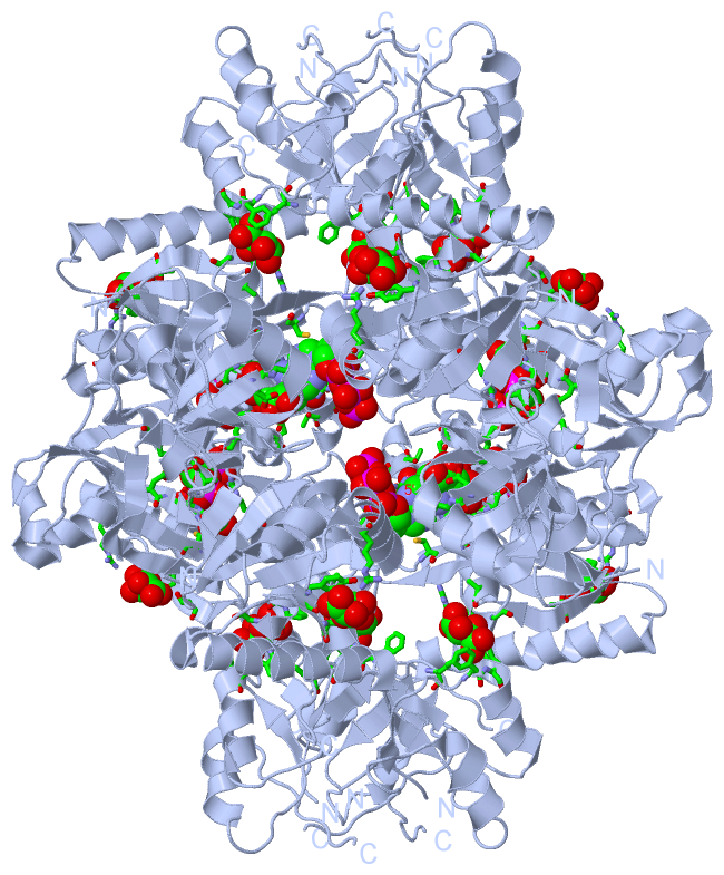
    GO:0003879    ATP phosphoribosyltransferase activity    Catalysis of the reaction: 1-(5-phospho-D-ribosyl)-ATP + diphosphate = ATP + 5-phospho-alpha-D-ribose 1-diphosphate.
    GO:0000287    magnesium ion binding    Interacting selectively and non-covalently with magnesium (Mg) ions.
    GO:0046872    metal ion binding    Interacting selectively and non-covalently with any metal ion.
    GO:0000166    nucleotide binding    Interacting selectively and non-covalently with a nucleotide, any compound consisting of a nucleoside that is esterified with (ortho)phosphate or an oligophosphate at any hydroxyl group on the ribose or deoxyribose.
    GO:0016740    transferase activity    Catalysis of the transfer of a group, e.g. a methyl group, glycosyl group, acyl group, phosphorus-containing, or other groups, from one compound (generally regarded as the donor) to another compound (generally regarded as the acceptor). Transferase is the systematic name for any enzyme of EC class 2.
    GO:0016757    transferase activity, transferring glycosyl groups    Catalysis of the transfer of a glycosyl group from one compound (donor) to another (acceptor).
biological process
    GO:0008652    cellular amino acid biosynthetic process    The chemical reactions and pathways resulting in the formation of amino acids, organic acids containing one or more amino substituents.
    GO:0000105    histidine biosynthetic process    The chemical reactions and pathways resulting in the formation of histidine, 2-amino-3-(1H-imidazol-4-yl)propanoic acid.
cellular component
    GO:0005737    cytoplasm    All of the contents of a cell excluding the plasma membrane and nucleus, but including other subcellular structures.
    GO:0005829    cytosol    The part of the cytoplasm that does not contain organelles but which does contain other particulate matter, such as protein complexes.

 Visualization

(-) Interactive Views

Asymmetric Unit
  Complete Structure
    Jena3D(integrated viewing of ligand, site, SAP, PROSITE, SCOP information)
    WebMol | AstexViewer[tm]@PDBe
(Java Applets, require no local installation except for Java; loading may be slow)
    STRAP
(Java WebStart application, automatic local installation, requires Java; full application with system access!)
    RasMol
(require local installation)
    Molscript (VRML)
(requires installation of a VRML viewer; select preferred view via VRML and generate a mono or stereo PDF format file)
 
  Ligands, Modified Residues, Ions
    PRT  [ RasMol | Jena3D ]  +environment [ RasMol | Jena3D ]
    TLA  [ RasMol | Jena3D ]  +environment [ RasMol | Jena3D ]
 
  Sites
    AC1  [ RasMol ]  +environment [ RasMol ]
    AC2  [ RasMol ]  +environment [ RasMol ]
    AC3  [ RasMol ]  +environment [ RasMol ]
 
  Cis Peptide Bonds
(no "Cis Peptide Bonds" information available for 1q1k)
 
Biological Unit
  Complete Structure
    Biological Unit 1  [ Jena3D ]

(-) Still Images

Jmol
  protein: cartoon or spacefill or dots and stick; nucleic acid: cartoon and stick; ligands: spacefill; active site: stick
Molscript
  protein, nucleic acid: cartoon; ligands: spacefill; active site: ball and stick

 Databases and Analysis Tools

(-) Databases

Access by PDB/NDB ID
  1q1k
    Family and Domain InformationProDom | SYSTERS
    General Structural InformationGlycoscienceDB | MMDB | NDB | OCA | PDB | PDBe | PDBj | PDBsum | PDBWiki | PQS | PROTEOPEDIA
    Orientation in MembranesOPM
    Protein SurfaceSURFACE
    Secondary StructureDSSP (structure derived) | HSSP (homology derived)
    Structural GenomicsGeneCensus
    Structural NeighboursCE | VAST
    Structure ClassificationCATH | Dali | SCOP
    Validation and Original DataBMRB Data View | BMRB Restraints Grid | EDS | PROCHECK | RECOORD | WHAT_CHECK
 
Access by UniProt ID/Accession number
  HIS1_ECOLI | P60757
    Comparative Protein Structure ModelsModBase
    Genomic InformationEnsembl
    Protein-protein InteractionDIP
    Sequence, Family and Domain InformationInterPro | Pfam | SMART | UniProtKB/SwissProt
 
Access by Enzyme Classificator   (EC Number)
  2.4.2.17
    General Enzyme InformationBRENDA | EC-PDB | Enzyme | IntEnz
    PathwayKEGG | MetaCyc
 
Access by Disease Identifier   (MIM ID)
  (no 'MIM ID' available)
    Disease InformationOMIM
 
Access by GenAge ID
  (no 'GenAge ID' available)
    Age Related InformationGenAge

(-) Analysis Tools

Access by PDB/NDB ID
    Domain InformationXDom
    Interatomic Contacts of Structural UnitsCSU
    Ligand-protein ContactsLPC
    Protein CavitiescastP
    Sequence and Secondary StructurePDBCartoon
    Structure AlignmentSTRAP(Java WebStart application, automatic local installation, requires Java; full application with system access!)
    Structure and Sequence BrowserSTING
 
Access by UniProt ID/Accession number
  HIS1_ECOLI | P60757
    Protein Disorder PredictionDisEMBL | FoldIndex | GLOBPLOT (for more information see DisProt)

 Related Entries

(-) Entries Sharing at Least One Protein Chain (UniProt ID)

UniProtKB/Swiss-Prot
        HIS1_ECOLI | P607571h3d

(-) Related Entries Specified in the PDB File

(no "Related Entries Specified in the PDB File" available for 1Q1K)