Show PDB file:   
         Plain Text   HTML   (compressed file size)
QuickSearch:   
by PDB,NDB,UniProt,PROSITE Code or Search Term(s)  
(-)Asymmetric Unit
(-)Asym. Unit - sites
(-)Biological Unit 1
(-)Biol. Unit 1 - sites
(-)Biological Unit 2
collapse expand < >
Image Asymmetric Unit
Asymmetric Unit  (Jmol Viewer)
Image Asym. Unit - sites
Asym. Unit - sites  (Jmol Viewer)
Image Biological Unit 1
Biological Unit 1  (Jmol Viewer)
Image Biol. Unit 1 - sites
Biol. Unit 1 - sites  (Jmol Viewer)
Image Biological Unit 2
Biological Unit 2  (Jmol Viewer)

(-) Description

Title :  CRYSTAL STRUCTURE OF JTOR68S
 
Authors :  C. Dealwis, V. Gupta, M. Wilkerson
Date :  30 Jun 03  (Deposition) - 17 Aug 04  (Release) - 24 Feb 09  (Revision)
Method :  X-RAY DIFFRACTION
Resolution :  1.90
Chains :  Asym. Unit :  A,B
Biol. Unit 1:  A  (1x)
Biol. Unit 2:  B  (1x)
Keywords :  Light Chain, Amyloidosis, Immune System (Keyword Search: [Gene Ontology, PubMed, Web (Google))
 
Reference :  J. S. Wall, V. Gupta, M. Wilkerson, M. Schell, R. Loris, P. Adams, A. Solomon, F. Stevens, C. Dealwis
Structural Basis Of Light Chain Amyloidogenicity: Comparison Of The Thermodynamic Properties, Fibrillogenic Potential And Tertiary Structural Features Of Four Vlambda6 Proteins.
J. Mol. Recog. V. 17 323 2004
PubMed-ID: 15227639  |  Reference-DOI: 10.1002/JMR.681
(for further references see the PDB file header)

(-) Compounds

Molecule 1 - IMMUNOGLOBULIN LAMBDA CHAIN VARIABLE REGION
    ChainsA, B
    EngineeredYES
    Expression SystemESCHERICHIA COLI
    Expression System PlasmidPET-27B
    Expression System Taxid562
    Expression System Vector TypePLASMID
    GeneJTO
    MutationYES
    Organism CommonHUMAN
    Organism ScientificHOMO SAPIENS
    Organism Taxid9606
    SynonymJTO

 Structural Features

(-) Chains, Units

  12
Asymmetric Unit AB
Biological Unit 1 (1x)A 
Biological Unit 2 (1x) B

Summary Information (see also Sequences/Alignments below)

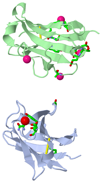
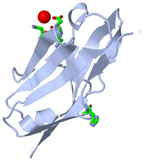
(-) Ligands, Modified Residues, Ions  (1, 4)

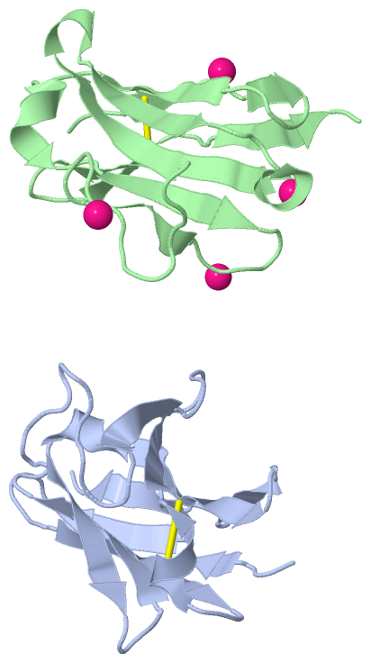
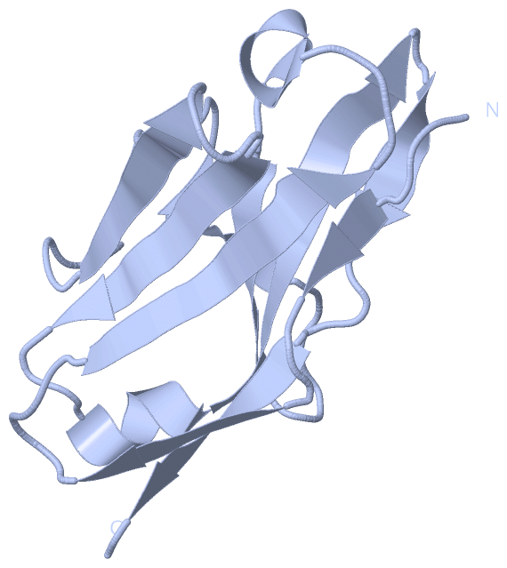
Asymmetric Unit (1, 4)
No.NameCountTypeFull Name
1CD4Ligand/IonCADMIUM ION
Biological Unit 1 (0, 0)
No.NameCountTypeFull Name
1CD-1Ligand/IonCADMIUM ION
Biological Unit 2 (0, 0)
No.NameCountTypeFull Name
1CD-1Ligand/IonCADMIUM ION

(-) Sites  (4, 4)

Asymmetric Unit (4, 4)
No.NameEvidenceResiduesDescription
1AC1SOFTWAREHIS A:8 , ALA B:43 , TYR B:87 , GLY B:100 , GLY B:101BINDING SITE FOR RESIDUE CD B 201
2AC2SOFTWARETYR A:49 , GLU A:50 , GLN A:53 , HOH A:136BINDING SITE FOR RESIDUE CD B 202
3AC3SOFTWARETHR B:80 , GLU B:81 , GLU B:83BINDING SITE FOR RESIDUE CD B 203
4AC4SOFTWAREGLY A:41 , GLN B:6 , PRO B:7 , HIS B:8BINDING SITE FOR RESIDUE CD B 204

(-) SS Bonds  (2, 2)

Asymmetric Unit
No.Residues
1A:23 -A:88
2B:23 -B:88

(-) Cis Peptide Bonds  (0, 0)

(no "Cis Peptide Bond" information available for 1PW3)

 Sequence-Structure Mapping

(-) SAPs(SNPs)/Variants  (0, 0)

(no "SAP(SNP)/Variant" information available for 1PW3)

(-) PROSITE Motifs  (0, 0)

(no "PROSITE Motif" information available for 1PW3)

(-) Exons   (0, 0)

(no "Exon" information available for 1PW3)

(-) Sequences/Alignments

Asymmetric Unit
   Reformat: Number of residues per line =  ('0' or empty: single-line sequence representation)
  Number of residues per labelling interval =   
  UniProt sequence: complete  aligned part    
   Show mapping: SCOP domains CATH domains Pfam domains Secondary structure (by author)
SAPs(SNPs) PROSITE motifs Exons
(details for a mapped element are shown in a popup box when the mouse pointer rests over it)
Chain A from PDB  Type:PROTEIN  Length:111
 aligned with Q96JD1_HUMAN | Q96JD1 from UniProtKB/TrEMBL  Length:112

    Alignment length:112
                                    10        20        30        40        50        60        70        80        90       100       110  
         Q96JD1_HUMAN     1 NFMLTQPHSVSESPGKTITISCTRSSGSIASNYVQWYQQRPGSAPTTVIYEDNQRPSGVPDRFSGSIDSSSNSASLTISGLKTEDEADYYCQSYDSNNYALFGGGTQLTVLG 112
               SCOP domains d1pw3a_ A: Immunoglobulin light chain lambda variable domain, VL-lambda                                          SCOP domains
               CATH domains 1pw3A00 A:1-108 Immunoglobulins                                                                                  CATH domains
               Pfam domains ---------------------------------------------------------------------------------------------------------------- Pfam domains
         Sec.struct. author ...ee...eeee.....eeeeeee...hhhhh..eeeee......eeee.............eeeeee....eeeeee...hhhhheeeeeeeee..e-eee...eeeee.. Sec.struct. author
                 SAPs(SNPs) ---------------------------------------------------------------------------------------------------------------- SAPs(SNPs)
                    PROSITE ---------------------------------------------------------------------------------------------------------------- PROSITE
                 Transcript ---------------------------------------------------------------------------------------------------------------- Transcript
                 1pw3 A   1 NFMLNQPHSVSESPGKTVTISCTRSSGNIDSNYVQWYQQRPGSAPITVIYEDNQRPSGVPDRFAGSIDSSSNSASLTISGLKTEDEADYYCQSYDARN-VVFGGGTRLTVLG 108
                                    11        21      ||29        39        49        59       |67        77        87       |96       106  
                                    9|              27A|                                     66A|                           95 |            
                                    11               27B                                      66B                             96            

Chain B from PDB  Type:PROTEIN  Length:111
 aligned with Q96JD1_HUMAN | Q96JD1 from UniProtKB/TrEMBL  Length:112

    Alignment length:112
                                    10        20        30        40        50        60        70        80        90       100       110  
         Q96JD1_HUMAN     1 NFMLTQPHSVSESPGKTITISCTRSSGSIASNYVQWYQQRPGSAPTTVIYEDNQRPSGVPDRFSGSIDSSSNSASLTISGLKTEDEADYYCQSYDSNNYALFGGGTQLTVLG 112
               SCOP domains d1pw3b_ B: Immunoglobulin light chain lambda variable domain, VL-lambda                                          SCOP domains
               CATH domains 1pw3B00 B:1-108 Immunoglobulins                                                                                  CATH domains
           Pfam domains (1) V-set-1pw3B01 B:1-106                                                                                         -- Pfam domains (1)
           Pfam domains (2) V-set-1pw3B02 B:1-106                                                                                         -- Pfam domains (2)
         Sec.struct. author ...ee...eeee.....eeeeeee...hhhhh..eeeee......eeee.............eeeeeehhh.eeeeee...hhhhheeeeeeee....-eee...eeeee.. Sec.struct. author
                 SAPs(SNPs) ---------------------------------------------------------------------------------------------------------------- SAPs(SNPs)
                    PROSITE ---------------------------------------------------------------------------------------------------------------- PROSITE
                 Transcript ---------------------------------------------------------------------------------------------------------------- Transcript
                 1pw3 B   1 NFMLNQPHSVSESPGKTVTISCTRSSGNIDSNYVQWYQQRPGSAPITVIYEDNQRPSGVPDRFAGSIDSSSNSASLTISGLKTEDEADYYCQSYDARN-VVFGGGTRLTVLG 108
                                    11        21      ||29        39        49        59       |67        77        87       |96       106  
                                    9|              27A|                                     66A|                           95 |            
                                    11               27B                                      66B                             96            

   Legend:   → Mismatch (orange background)
  - → Gap (green background, '-', border residues have a numbering label)
    → Modified Residue (blue background, lower-case, 'x' indicates undefined single-letter code, labelled with number + name)
  x → Chemical Group (purple background, 'x', labelled with number + name, e.g. ACE or NH2)
  extra numbering lines below/above indicate numbering irregularities and modified residue names etc., number ends below/above '|'

 Classification and Annotation

(-) SCOP Domains  (1, 2)

Asymmetric Unit

(-) CATH Domains  (1, 2)

Asymmetric Unit
(-)
Class: Mainly Beta (13760)

(-) Pfam Domains  (1, 2)

Asymmetric Unit
(-)
Clan: Ig (577)

(-) Gene Ontology  (0, 0)

Asymmetric Unit(hide GO term definitions)
    (no "Gene Ontology" information available for 1PW3)

 Visualization

(-) Interactive Views

Asymmetric Unit
  Complete Structure
    Jena3D(integrated viewing of ligand, site, SAP, PROSITE, SCOP information)
    WebMol | AstexViewer[tm]@PDBe
(Java Applets, require no local installation except for Java; loading may be slow)
    STRAP
(Java WebStart application, automatic local installation, requires Java; full application with system access!)
    RasMol
(require local installation)
    Molscript (VRML)
(requires installation of a VRML viewer; select preferred view via VRML and generate a mono or stereo PDF format file)
 
  Ligands, Modified Residues, Ions
    CD  [ RasMol | Jena3D ]  +environment [ RasMol | Jena3D ]
 
  Sites
    AC1  [ RasMol ]  +environment [ RasMol ]
    AC2  [ RasMol ]  +environment [ RasMol ]
    AC3  [ RasMol ]  +environment [ RasMol ]
    AC4  [ RasMol ]  +environment [ RasMol ]
 
  Cis Peptide Bonds
(no "Cis Peptide Bonds" information available for 1pw3)
 
Biological Units
  Complete Structure
    Biological Unit 1  [ Jena3D ]
    Biological Unit 2  [ Jena3D ]

(-) Still Images

Jmol
  protein: cartoon or spacefill or dots and stick; nucleic acid: cartoon and stick; ligands: spacefill; active site: stick
Molscript
  protein, nucleic acid: cartoon; ligands: spacefill; active site: ball and stick

 Databases and Analysis Tools

(-) Databases

Access by PDB/NDB ID
  1pw3
    Family and Domain InformationProDom | SYSTERS
    General Structural InformationGlycoscienceDB | MMDB | NDB | OCA | PDB | PDBe | PDBj | PDBsum | PDBWiki | PQS | PROTEOPEDIA
    Orientation in MembranesOPM
    Protein SurfaceSURFACE
    Secondary StructureDSSP (structure derived) | HSSP (homology derived)
    Structural GenomicsGeneCensus
    Structural NeighboursCE | VAST
    Structure ClassificationCATH | Dali | SCOP
    Validation and Original DataBMRB Data View | BMRB Restraints Grid | EDS | PROCHECK | RECOORD | WHAT_CHECK
 
Access by UniProt ID/Accession number
  Q96JD1_HUMAN | Q96JD1
    Comparative Protein Structure ModelsModBase
    Genomic InformationEnsembl
    Protein-protein InteractionDIP
    Sequence, Family and Domain InformationInterPro | Pfam | SMART | UniProtKB/TrEMBL
 
Access by Enzyme Classificator   (EC Number)
  (no 'Enzyme Classificator' available)
    General Enzyme InformationBRENDA | EC-PDB | Enzyme | IntEnz
    PathwayKEGG | MetaCyc
 
Access by Disease Identifier   (MIM ID)
  (no 'MIM ID' available)
    Disease InformationOMIM
 
Access by GenAge ID
  (no 'GenAge ID' available)
    Age Related InformationGenAge

(-) Analysis Tools

Access by PDB/NDB ID
    Domain InformationXDom
    Interatomic Contacts of Structural UnitsCSU
    Ligand-protein ContactsLPC
    Protein CavitiescastP
    Sequence and Secondary StructurePDBCartoon
    Structure AlignmentSTRAP(Java WebStart application, automatic local installation, requires Java; full application with system access!)
    Structure and Sequence BrowserSTING
 
Access by UniProt ID/Accession number
  Q96JD1_HUMAN | Q96JD1
    Protein Disorder PredictionDisEMBL | FoldIndex | GLOBPLOT (for more information see DisProt)

 Related Entries

(-) Entries Sharing at Least One Protein Chain (UniProt ID)

UniProtKB/Swiss-Prot
        Q96JD1_HUMAN | Q96JD13bdx
UniProtKB/TrEMBL
        Q96JD1_HUMAN | Q96JD15c9k

(-) Related Entries Specified in the PDB File

1cdo 1pew