|
|
|
|
 Description
|
|
Compounds
|
||||||||||||||||||||||||||||||||||||
Chains, Units
Summary Information (see also Sequences/Alignments below) |
Ligands, Modified Residues, Ions (0, 0)| (no "Ligand,Modified Residues,Ions" information available for 1PUZ) |
Sites (0, 0)| (no "Site" information available for 1PUZ) |
SS Bonds (0, 0)| (no "SS Bond" information available for 1PUZ) |
Cis Peptide Bonds (0, 0)| (no "Cis Peptide Bond" information available for 1PUZ) |
SAPs(SNPs)/Variants (0, 0)| (no "SAP(SNP)/Variant" information available for 1PUZ) |
PROSITE Motifs (0, 0)| (no "PROSITE Motif" information available for 1PUZ) |
Exons (0, 0)| (no "Exon" information available for 1PUZ) |
Sequences/Alignments
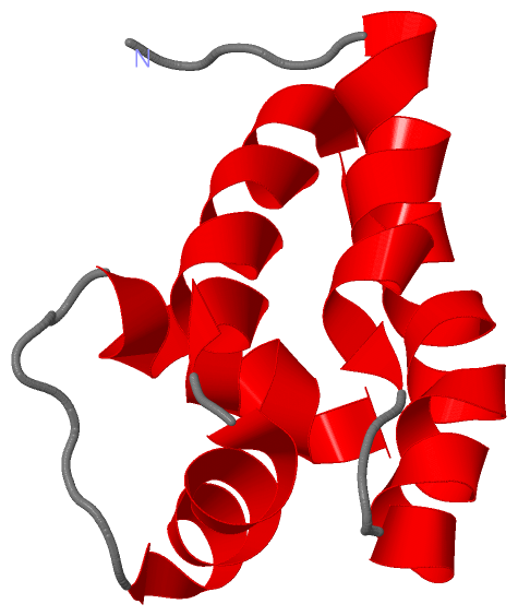
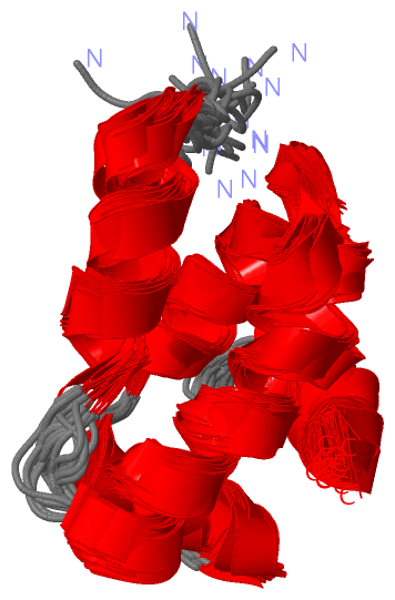
NMR StructureChain A from PDB Type:PROTEIN Length:82 aligned with Q7DDK1_NEIMB | Q7DDK1 from UniProtKB/TrEMBL Length:82 Alignment length:82 10 20 30 40 50 60 70 80 Q7DDK1_NEIMB 1 MMVFDDIAKRKIRFQTRRGLLELDLIFGRFMEKEFEHLSDKELSEFSEILEFQDQELLALINGHSETDKGHLIPMLEKIRRA 82 SCOP domains d1puza_ A: Hypothetical protein NMA1147 SCOP domains CATH domains 1puzA00 A:1-82 Ygfy CATH domains Pfam domains -------------Sdh5-1puzA01 A:14-63 ------------------- Pfam domains SAPs(SNPs) ---------------------------------------------------------------------------------- SAPs(SNPs) PROSITE ---------------------------------------------------------------------------------- PROSITE Transcript ---------------------------------------------------------------------------------- Transcript 1puz A 1 MMVFDDIAKRKIRFQTRRGLLELDLIFGRFMEKEFEHLSDKELSEFSEILEFQDQELLALINGHSETDKGHLIPMLEKIRRA 82 10 20 30 40 50 60 70 80
|
||||||||||||||||||||
SCOP Domains (1, 1)
NMR Structure
 |
CATH Domains (1, 1)
NMR Structure
|
Pfam Domains (1, 1)
NMR Structure
|
Gene Ontology (0, 0)|
NMR Structure(hide GO term definitions)
(no "Gene Ontology" information available for 1PUZ)
|
Interactive Views
|
||||||||||||||||||||||||||||||||||||||||||||||||||||||||||||||||||||||||||||||||||||||||||||||||||||||||||||||||||||
Still Images
|
||||||||||||||||
Databases
|
||||||||||||||||||||||||||||||||||||||||||||||||||||||||||||||||||||||||||||||||||||||||||||||||||||||||||||||||||||||||||||||||||||||||||||||||||||||||||||||||
Analysis Tools
|
|||||||||||||||||||||||||||||||||||||||||||||||||||||||||||||
Entries Sharing at Least One Protein Chain (UniProt ID)
Related Entries Specified in the PDB File
|
|